




Tratamiento del sarcoma uterino
Reciba una atención excepcional para el sarcoma uterino y otros tipos de cáncer ginecológico en Montefiore Einstein Comprehensive Cancer Center. Nuestro equipo multidisciplinario cuenta con una amplia experiencia en el tratamiento de este tipo de cáncer. Somos uno de los primeros centros del cáncer designados por el NCI y, como tal, llevamos más de 50 años liderando en la investigación, el diagnóstico y el tratamiento de más de 200 tipos de cáncer.
Estamos a la vanguardia del tratamiento y la investigación del sarcoma uterino y otros cánceres ginecológicos. Ofrecemos planes de tratamiento integrales y personalizados según las necesidades de nuestras pacientes. En el Montefiore Einstein Comprehensive Cancer Center, ponemos a su disposición una atención innovadora y avanzada basada en las últimas investigaciones sobre el cáncer, incluido el acceso a ensayos clínicos.
Cuando necesite atención para el sarcoma uterino, recurra a nuestros proveedores, a quienes les apasiona erradicar el cáncer y abordar todas sus necesidades de salud.

El Montefiore Einstein Comprehensive Cancer Center, designado como centro integral del cáncer por el National Cancer Institute (NCI), apoya la misión y las normas del NCI. La siguiente información sobre los tipos de cáncer, prevención y tratamientos ha sido facilitada por el NCI.
Tratamiento del sarcoma uterino (PDQ®): versión para pacientes
Información general sobre el sarcoma uterino
Puntos clave
- El sarcoma uterino es una enfermedad en la que se forman células malignas (cancerosas) en los músculos del útero u otros tejidos que sostienen el útero.
- Los tratamientos anteriores con radioterapia en la pelvis pueden aumentar el riesgo de sarcoma uterino.
- Uno de los signos del sarcoma uterino es sangrado anómalo.
- Para diagnosticar el sarcoma uterino se utilizan pruebas que examinan el útero.
- Hay ciertos factores que afectan al pronóstico (probabilidad de recuperación) y a las opciones de tratamiento.
El sarcoma uterino es una enfermedad en la que se forman células malignas (cancerosas) en los músculos del útero u otros tejidos que sostienen el útero.
El útero es parte del sistema reproductor femenino. El útero es un órgano hueco con forma de pera que se encuentra en la pelvis, donde crece el feto. El cuello uterino se encuentra en el extremo inferior y estrecho del útero y conduce hasta la vagina.
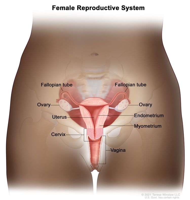
Anatomía del aparato reproductor femenino. Entre los órganos del aparato reproductor femenino están el útero, los ovarios, las trompas de Falopio, el cuello uterino y la vagina. El útero tiene una capa muscular externa, llamada miometrio, y una capa de tejido interno que lo reviste, llamada endometrio.
El sarcoma uterino es un tipo de cáncer muy poco común que se forma en los músculos uterinos o en los tejidos que lo sostienen. Para obtener más información sobre otros tipos de sarcomas, visite Tratamiento del sarcoma de tejido blando.
El sarcoma uterino es diferente del cáncer de endometrio, una enfermedad en la que el cáncer se forma en el tejido que recubre el útero. El carcinosarcoma es un subtipo de cáncer de endometrio y se estadifica según las definiciones de cáncer de endometrio. Para más información, visite Tratamiento del cáncer de endometrio.
Los sarcomas uterinos pueden ser leiomiosarcomas, sarcomas del estroma endometrial o adenosarcomas.
Los tratamientos anteriores con radioterapia en la pelvis pueden aumentar el riesgo de sarcoma uterino.
Cualquier factor que aumente la probabilidad de desarrollar una enfermedad se denomina factor de riesgo. No todas las personas con uno o más de estos factores de riesgo desarrollarán cáncer testicular, y también se desarrollará en algunas personas que no tienen ningún factor de riesgo conocido. Hable con su médico si cree que puede estar en riesgo.
- Tratamientos anteriores con radioterapia en la pelvis
- Tratamientos con tamoxifeno para el cáncer de mama. Si toma este medicamento, hágase un examen pélvico anualmente e informe sobre cualquier sangrado vaginal (excepto el menstrual) lo antes posible.
Uno de los signos del sarcoma uterino es sangrado anómalo.
El sangrado vaginal anómalo y otros signos y síntomas pueden deberse a un sarcoma uterino o a otras afecciones. Consulte con su médico si presenta:
- Sangrado que no forma parte de los períodos menstruales
- Sangrado después de la menopausia
- Una masa en la vagina
- Dolor o sensación de estar llena en el abdomen
- Micción frecuente
Para diagnosticar el sarcoma uterino se utilizan pruebas que examinan el útero.
Además de preguntarle sobre su historial de salud personal y familiar y de hacerle un reconocimiento físico, es posible que el médico realice las siguientes pruebas y procedimientos:
- Examen pélvico: un examen de la vagina, cuello uterino, útero, trompas de Falopio, ovarios, y las recto. Un espéculo se introduce en la vagina y el médico o enfermero examina la vagina y el cuello uterino en busca de signos de enfermedad. Un Prueba de Papanicolaou del cuello uterino por lo general se hace. El médico o enfermero también inserta uno o dos lubricadosdedos, con guantes y lubricados, dentro de la vagina y coloca la otra mano sobre la parte inferior del abdomen para examinar el tamaño, la forma y la posición del útero y los ovarios. El médico o enfermero también introducen un dedo, cubierto por un guante y lubricado, en el recto para palpar si hay bultos o zonas anómalas.
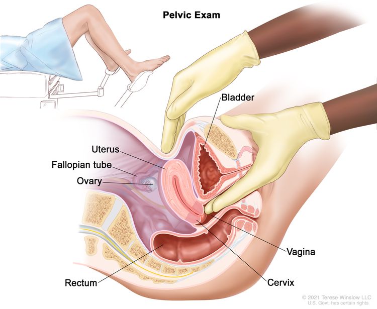
Examen pélvico. Un médico o enfermero introduce uno o dos dedos de una mano, con guantes y lubricados, en la vagina, mientras presiona la parte inferior del abdomen con la otra mano. Esto se hace para palpar el tamaño, la forma y la posición del útero y los ovarios. También se examinan la vagina, el cuello uterino, las trompas de Falopio y el recto.
- Prueba de Papanicolaou: procedimiento para recolectar células de la superficie del cuello uterino y la vagina. Se utiliza un trozo de algodón, un cepillo o una espátula pequeña de madera para hacer un raspado suave de las células del cuello uterino y la vagina. Las células se observan con microscopio para saber si son anómalas. Este procedimiento también se llama prueba de Papanicolaou. Debido a que el sarcoma uterino comienza dentro del útero, es posible que este cáncer no aparezca en la prueba de Papanicolaou.
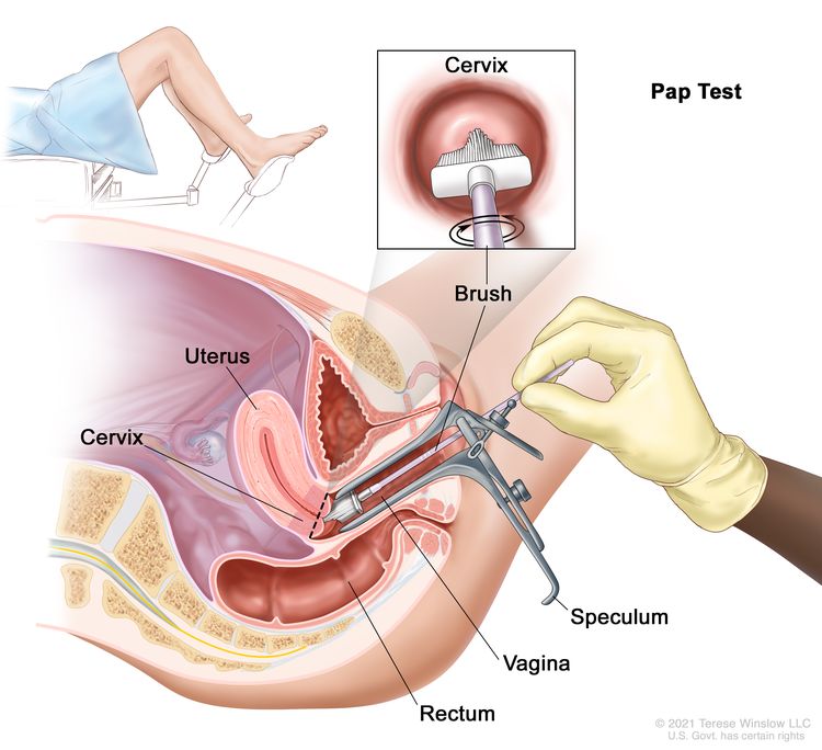
Prueba de Pap. Se introduce un espéculo en la vagina para ensancharla. A continuación, se introduce un cepillo para recolectar células del cuello uterino. Las células se examinan con microscopio para detectar signos de enfermedad.
- Examen de ultrasonido transvaginal: procedimiento utilizado para examinar la vagina, el útero, las trompas de Falopio y la vejiga. Un transductor de ultrasonido (sonda) se inserta en la vagina y se usa para hacer rebotar ondas sonoras de alta energía (ecografía) de los tejidos u órganos internos y hacer ecos. Los ecos forman una imagen de los tejidos del cuerpo llamada ecograma. El médico puede identificar tumores observando la ecografía.
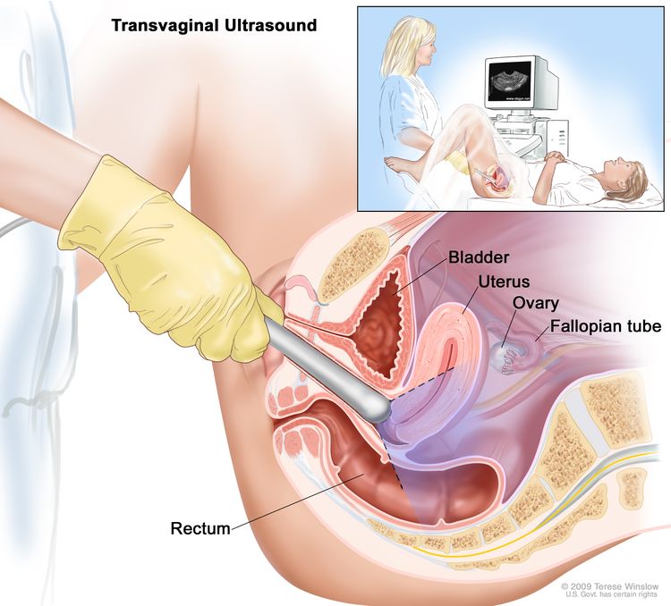
Ecografía transvaginal. Se introduce en la vagina una sonda de ecografía conectada a una computadora y se mueve suavemente para visualizar diferentes órganos. La sonda emite ondas sonoras en los órganos y tejidos internos para generar ecos que forman un ecograma (imagen computarizada).
- Dilatación y curetaje: procedimiento para extraer muestras de tejido del revestimiento interno del útero. El cuello uterino es dilatado y una cureta (instrumento en forma de cuchara) se inserta en el útero para extraer tejido. Las muestras de tejido se examinan con un microscopio para detectar signos de enfermedad. Este procedimiento también se llama D y C.
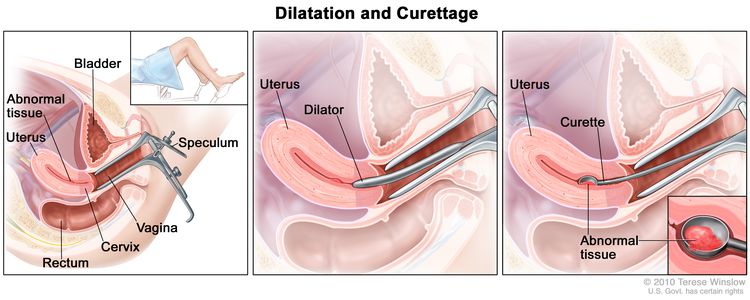
Dilatación y curetaje (D y C). Se inserta un espéculo en la vagina para ensancharla y observar el cuello uterino (primer gráfico). Se utiliza un dilatador para ensanchar el cuello uterino (gráfico del medio). Se coloca una cureta a través del cuello uterino hasta el útero para raspar el tejido anómalo (último gráfico).
- Biopsia endometrial: consiste en la extracción de tejido del endometrio (revestimiento interno del útero) mediante la inserción de un tubo delgado y flexible a través del cuello uterino hasta el interior del útero. El tubo se utiliza para raspar suavemente una pequeña cantidad de tejido del endometrio y luego extraer las muestras. Un patólogo observa el tejido con microscopio para detectar células cancerosas.
Hay ciertos factores que afectan al pronóstico (probabilidad de recuperación) y a las opciones de tratamiento.
El pronóstico y las opciones de tratamiento dependen de los siguientes factores:
- El estadio del cáncer
- El tipo y tamaño del tumor
- La salud general del paciente
- Si el cáncer acaba de ser diagnosticado o ha reaparecido (regresado)
Estadios del sarcoma uterino
Puntos clave
- Una vez que se diagnostica el sarcoma uterino, se realizan pruebas para determinar si las células cancerosas se han extendido dentro del útero o hacia otras partes del cuerpo.
- El sarcoma uterino se puede diagnosticar, clasificar por estadios y tratar en la misma cirugía.
- El cáncer se propaga por el cuerpo de tres maneras.
- El cáncer puede extenderse desde donde comenzó a otras partes del cuerpo.
- Para los leiomiosarcomas y los sarcomas del estroma endometrial se utiliza el siguiente sistema de estadificación FIGO:
- Estadio I
- Estadio II
- Estadio III
- Estadio IV
- Para los adenosarcomas se utiliza el siguiente sistema de estadificación FIGO:
- Estadio I
- Estadio II
- Estadio III
- Estadio IV
- El sarcoma uterino puede recidivar (volver) después de haber sido tratado.
Una vez que se diagnostica el sarcoma uterino, se realizan pruebas para determinar si las células cancerosas se han extendido dentro del útero o hacia otras partes del cuerpo.
El proceso que se utiliza para determinar si el cáncer se ha extendido dentro del útero o a otras partes del cuerpo se denomina estadificación. La información obtenida del proceso de estadificación determina el estadio de la enfermedad. Es importante conocer el estadio para planificar el tratamiento. En el proceso de estadificación se pueden utilizar los siguientes procedimientos:
- Estudio bioquímico de la sangre: procedimiento en el que se analiza una muestra de sangre para medir las cantidades de ciertas sustancias liberadas a la sangre por los órganos y tejidos del cuerpo. Una cantidad inusual (mayor o menor de lo normal) de una sustancia puede ser un signo de enfermedad.
- Análisis de CA-125: prueba que mide el nivel de CA-125 en la sangre. El CA-125 es una sustancia que las células liberan al torrente sanguíneo. Un nivel elevado de CA-125 a veces es un signo de cáncer u otra afección.
- Radiografía de tórax: radiografía de los órganos y huesos del interior del tórax. Un rayo X es un tipo de haz de energía que puede atravesar el cuerpo y plasmarse en una película, generando una imagen de áreas internas del cuerpo.
- Examen de ultrasonido transvaginal: procedimiento utilizado para examinar la vagina, útero, trompas de Falopio, y las vejiga. Un transductor de ultrasonido (sonda) se inserta en la vagina y se usa para hacer rebotar ondas sonoras de alta energía (ecografía) de los tejidos u órganos internos y hacer ecos. Los ecos forman una imagen de los tejidos del cuerpo llamada ecograma. El médico puede identificar tumores observando la ecografía.
Ecografía transvaginal. Se introduce en la vagina una sonda de ecografía conectada a una computadora y se mueve suavemente para visualizar diferentes órganos. La sonda emite ondas sonoras en los órganos y tejidos internos para generar ecos que forman un ecograma (imagen computarizada).
- Exploración por tomografía computarizada (TAC): procedimiento que genera una serie de imágenes detalladas de zonas internas del cuerpo, como el abdomen y la pelvis, tomadas desde diferentes ángulos. Las imágenes son tomadas por una computadora conectada a una máquina de rayos X. Se puede inyectar un tinte en una vena o ingerirlo para ayudar a que los órganos o tejidos se vean más claramente. Este procedimiento también se llama tomografía computarizada, tomografía computerizada o tomografía axial computarizada.
- Cistoscopia: procedimiento para observar el interior de la vejiga y uretra para buscar anómalas zonas. Un cistoscopio se introduce a través de la uretra hasta la vejiga. Un cistoscopio es un instrumento delgado en forma de tubo, con una luz y una lente para observar. A veces tiene una herramienta para extraer muestras de tejido que se observan con microscopio para detectar signos de cáncer.
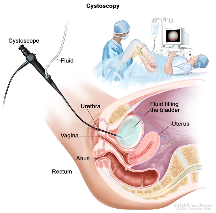
Cistoscopia. Se introduce un cistoscopio (instrumento delgado en forma de tubo, con una luz y una lente para observar) a través de la uretra hasta la vejiga. Se usa un líquido para llenar la vejiga. El médico observa una imagen de la pared interna de la vejiga en el monitor de una computadora para verificar si hay zonas anómalas.
El sarcoma uterino se puede diagnosticar, clasificar por estadios y tratar en la misma cirugía.
La cirugía se utiliza para diagnosticar, estadificar y tratar el sarcoma uterino. Durante esta cirugía, el médico extirpa la mayor cantidad posible de cáncer. Se pueden utilizar los siguientes procedimientos para diagnosticar, estadificar y tratar el sarcoma uterino:
- Laparotomía: procedimiento quirúrgico en el que se realiza una incisión (corte) en la pared del abdomen para examinar el interior del abdomen en busca de signos de enfermedad. El tamaño de la incisión depende del motivo por el que se realiza la laparotomía. A veces se extraen órganos o se toman muestras de tejido y se examinan con microscopio para detectar signos de enfermedad.
- Lavados abdominales y pélvicos: procedimiento en el que se coloca una solución salina en las cavidades del cuerpo abdominal y pélvico. Al poco tiempo, se extrae el líquido y se observa con microscopio para detectar células cancerosas.
- Histerectomía abdominal total: procedimiento quirúrgico para extirpar el útero y cuello uterino a través de una gran incisión (corte) en el abdomen.
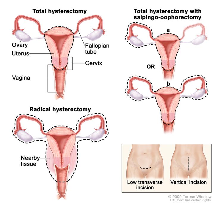
Histerectomía. El útero se extirpa quirúrgicamente con o sin otros órganos o tejidos. En una histerectomía total, se extirpan el útero y el cuello uterino. En una histerectomía total con salpingooforectomía, (a) se extirpan el útero, un ovario y una trompa de Falopio (unilaterales) o (b) se extirpan el útero, ambos ovarios y trompas de Falopio (bilaterales). En una histerectomía radical, se extirpan el útero, el cuello uterino, ambos ovarios, ambas trompas de Falopio y el tejido circundante. Estos procedimientos se realizan mediante una incisión transversal baja o una incisión vertical.
- Salpingooforectomía bilateral: cirugía para extirpar ambos ovarios y ambas trompas de Falopio.
- Linfadenectomía: procedimiento quirúrgico en el que se extirpan los ganglios linfáticos y se examinan con microscopio para detectar signos de cáncer. En el caso de la linfadenectomía regional, se extirpan algunos de los ganglios linfáticos de la zona del tumor. En el caso de la linfadenectomía radical, se extirpan la mayoría o todos los ganglios linfáticos de la zona del tumor. Este procedimiento también se llama disección de ganglios linfáticos.
Además de la cirugía, puede administrarse un tratamiento, tal como se describe en la sección Aspectos generales de las opciones de tratamiento de este resumen.
El cáncer se propaga por el cuerpo de tres maneras.
El cáncer puede extenderse desde donde comenzó a otras partes del cuerpo.
Cuando el cáncer se extiende a otra parte del cuerpo se denomina metástasis. Las células cancerosas se desprenden de donde comenzaron (tumor primario) y viajan a través del sistema linfático o la sangre.
- Sistema linfático: el cáncer entra en el sistema linfático, viaja a través de los vasos linfáticos y forma un tumor (tumor metastásico) en otra parte del cuerpo.
- Sangre: el cáncer llega a la sangre, viaja a través de los vasos sanguíneos y forma un tumor (tumor metastásico) en otra parte del cuerpo.
El tumor metastásico es el mismo tipo de cáncer que el tumor primario. Por ejemplo, si el sarcoma uterino se extiende al pulmón, las células cancerosas del pulmón son en realidad células de sarcoma uterino. La enfermedad es un sarcoma uterino metastásico, no un cáncer de pulmón.
El cáncer puede extenderse a través de los tejidos circundantes, el sistema linfático y la sangre:
- Tejidos: el cáncer se extiende desde el lugar donde comenzó y crece hacia las zonas circundantes.
- Sistema linfático: el cáncer se extiende desde el lugar donde comenzó hacia el sistema linfático. El cáncer viaja a través de los vasos linfáticos a otras partes del cuerpo.
- Sangre: el cáncer se extiende desde el lugar donde comenzó hacia la sangre. El cáncer viaja a través de los vasos sanguíneos a otras partes del cuerpo.
Para los leiomiosarcomas y los sarcomas del estroma endometrial se utiliza el siguiente sistema de estadificación FIGO:
Estadio I
En el estadio I, el tumor se encuentra únicamente en el útero. El estadio I se divide en estadios IA y IB:
- En el estadio IA, el tumor mide 5 centímetros o menos.
- En el estadio IB, el tumor mide más de 5 centímetros.

El tamaño de los tumores a menudo se mide en centímetros (cm) o pulgadas (in). A veces se usan alimentos comunes para mostrar el tamaño de un tumor en centímetros: una arveja o guisante (1 cm), un maní o cacahuate (2 cm), una uva (3 cm), una nuez (4 cm), una lima o limón verde (5 cm o 2 in), un huevo (6 cm), un durazno (7 cm) y un pomelo (10 cm o 4 in).
Estadio II
En el estadio II, el tumor se ha extendido más allá del útero pero no más allá de la pelvis. El estadio II se divide en estadios IIA y IIB:
- En el estadio IIA, el tumor se ha extendido al ovario, las trompas de Falopio o los tejidos conectivos alrededor del útero.
- En el estadio IIB, el tumor se ha extendido a otros tejidos de la pelvis.
Estadio III
En el estadio III, el tumor se ha extendido a los tejidos del abdomen. El estadio III se divide en estadios IIIA, IIIB y IIIC:
- En el estadio IIIA, el tumor se ha diseminado a un sitio en el abdomen.
- En el estadio IIIB, el tumor se ha diseminado a más de un sitio en el abdomen.
- En el estadio IIIC, el tumor se ha extendido a los ganglios linfáticos de la pelvis y/o alrededor de la aorta abdominal (el vaso sanguíneo más grande del abdomen).
Estadio IV
El estadio IV se divide en estadios IVA y IVB:
Para los adenosarcomas se utiliza el siguiente sistema de estadificación FIGO:
Estadio I
En el estadio I, el tumor se encuentra únicamente en el útero. El estadio I se divide en estadios IA, IB y IC:
- En el estadio IA, el tumor se encuentra en el endometrio o endocérvix (la parte interna del cuello uterino que forma un canal que conecta la vagina con el útero).
- En el estadio IB, el tumor se ha extendido hasta la mitad o menos del miometrio (la capa muscular externa del útero).
- En el estadio IC, el tumor se ha diseminado más allá de la mitad del espesor del miometrio.
Estadio II
En el estadio II, el tumor se ha extendido fuera del útero hacia la pelvis. Es estadio II se divide en estadio IIA y IIB:
- En el estadio IIA, el tumor se ha extendido al ovario, las trompas de Falopio o los tejidos conectivos alrededor del útero.
- En el estadio IIB, el tumor se ha extendido a otros tejidos de la pelvis.
Estadio III
En el estadio III, el tumor se ha extendido a los tejidos del abdomen. El estadio III se divide en estadios IIIA, IIIB y IIIC:
- En el estadio IIIA, el tumor se ha diseminado a un sitio en el abdomen.
- En el estadio IIIB, el tumor se ha diseminado a más de un sitio en el abdomen.
- En el estadio IIIC, el tumor se ha extendido a los ganglios linfáticos de la pelvis y/o alrededor de la aorta abdominal (el vaso sanguíneo más grande del abdomen).
Estadio IV
El estadio IV se divide en estadios IVA y IVB:
El sarcoma uterino puede recidivar (volver) después de haber sido tratado.
El cáncer puede reaparecer en el útero, la pelvis o en otras partes del cuerpo.
Descripción general de las opciones de tratamiento
Puntos clave
- Existen diferentes tipos de tratamiento para pacientes con sarcoma uterino.
- Se utilizan los siguientes tipos de tratamiento:
- Cirugía
- Radioterapia
- Quimioterapia
- Terapia hormonal
- Se están probando nuevos tipos de tratamiento en ensayos clínicos.
- El tratamiento para el sarcoma uterino puede causar efectos secundarios.
- Los pacientes pueden evaluar la posibilidad de participar en un ensayo clínico.
- Los pacientes pueden participar en ensayos clínicos antes, durante o después de comenzar el tratamiento contra el cáncer.
- Es posible que se necesiten cuidados de seguimiento.
Existen diferentes tipos de tratamiento para pacientes con sarcoma uterino.
Existen diferentes tipos de tratamientos disponibles para pacientes con sarcoma uterino. Algunos tratamientos son estándar (el tratamiento que se usa actualmente) y otros se están probando en ensayos clínicos. Un ensayo clínico de tratamiento es un estudio de investigación cuyo objetivo es ayudar a mejorar los tratamientos actuales u obtener información sobre nuevos tratamientos para pacientes con cáncer. Cuando los ensayos clínicos muestran que un nuevo tratamiento es mejor que el tratamiento estándar, el nuevo tratamiento puede convertirse en el tratamiento estándar. Es posible que las pacientes quieran pensar en participar en un ensayo clínico. Algunos ensayos clínicos están abiertos solo a pacientes que no han comenzado el tratamiento.
Se utilizan los siguientes tipos de tratamiento:
Cirugía
La cirugía es el tratamiento más común para el sarcoma uterino, como se describe en la sección Estadios del sarcoma uterino de este resumen.
Después de que el médico extirpe todo el cáncer que se puede observar en el momento de la cirugía, algunos pacientes podrían recibir quimioterapia o radioterapia para eliminar cualquier célula cancerosa restante. El tratamiento que se administra después de la cirugía para reducir el riesgo de reaparición del cáncer se denomina terapia adyuvante.
Radioterapia
La radioterapia es un tratamiento contra el cáncer que utiliza rayos X de alta energía u otros tipos de radiación para matar las células cancerosas o evitar que crezcan. Hay dos tipos de radioterapia:
- La radioterapia externa utiliza una máquina que envía radiación desde el exterior del cuerpo hacia la zona donde se encuentra el cáncer.
- La radioterapia interna utiliza una sustancia radiactiva sellada en agujas, semillas, cables o catéteres que se colocan directamente en el cáncer o cerca de él.
La forma en que se administra la radioterapia depende del tipo y estadio del cáncer que se está tratando. La radioterapia externa e interna se usa para tratar el sarcoma uterino y también puede usarse como terapia paliativa para aliviar los síntomas y mejorar la calidad de vida.
Quimioterapia
La quimioterapia es un tratamiento contra el cáncer que utiliza medicamentos para detener el crecimiento de las células cancerosas, ya sea destruyéndolas o impidiendo su división. Cuando la quimioterapia se administra por vía oral o se inyecta en una vena o músculo, los medicamentos entran en el torrente sanguíneo y pueden llegar a las células cancerosas de todo el cuerpo (quimioterapia sistémica).
Terapia hormonal
La terapia hormonal es un tratamiento contra el cáncer que elimina hormonas o bloquea su acción y detiene el crecimiento de las células cancerosas. Las hormonas son sustancias producidas por glándulas del cuerpo que circulan por el torrente sanguíneo. Algunas hormonas pueden provocar el crecimiento de ciertos cánceres. Si las pruebas muestran que las células cancerosas tienen lugares donde las hormonas pueden unirse (receptores), se utilizan medicamentos, cirugía o radioterapia para reducir la producción de hormonas o bloquear su funcionamiento.
Se están probando nuevos tipos de tratamiento en ensayos clínicos.
La información sobre los ensayos clínicos está disponible en el sitio web del NCI.
El tratamiento para el sarcoma uterino puede causar efectos secundarios.
Para obtener información sobre los efectos secundarios causados por el tratamiento para el cáncer, visite la página de efectos secundarios.
Los pacientes pueden evaluar la posibilidad de participar en un ensayo clínico.
Para algunos pacientes, participar en un ensayo clínico puede ser la mejor opción de tratamiento. Los ensayos clínicos son parte del proceso de investigación del cáncer y se realizan para determinar si los nuevos tratamientos para el cáncer son seguros y eficaces o mejores que el tratamiento estándar.
Muchos de los tratamientos estándar actuales para el cáncer se basan en ensayos clínicos anteriores. Los pacientes que participan en un ensayo clínico pueden recibir el tratamiento estándar o ser de los primeros en recibir uno nuevo.
Los pacientes que participan en ensayos clínicos también ayudan a mejorar la forma en que se tratará el cáncer en el futuro. Incluso cuando los ensayos clínicos no conducen a nuevos tratamientos efectivos, suelen responder a preguntas clave y contribuir de forma significativa al avance de la investigación.
Los pacientes pueden participar en ensayos clínicos antes, durante o después de comenzar el tratamiento contra el cáncer.
Algunos ensayos clínicos solo incluyen a pacientes que aún no han recibido tratamiento. Otros ensayos prueban tratamientos para pacientes cuyo cáncer no ha mejorado. También hay ensayos clínicos que prueban nuevas formas de evitar que el cáncer recidive (regrese) o de reducir los efectos secundarios del tratamiento del cáncer.
Se están realizando ensayos clínicos en muchas partes del país. Puede encontrar información sobre los ensayos clínicos respaldados por el NCI en el sitio web de búsqueda de ensayos clínicos del NCI. Puede encontrar ensayos clínicos respaldados por otras organizaciones en el sitio web ClinicalTrials.gov.
Es posible que se necesiten cuidados de seguimiento.
A medida que avanza el tratamiento, se le realizarán pruebas o controles de seguimiento. Es posible que se repitan algunas pruebas para diagnosticar o estadificar el cáncer con el fin de evaluar cómo está funcionando el tratamiento. Las decisiones sobre si continuar, modificar o suspender el tratamiento pueden basarse en los resultados de estas pruebas.
Algunas pruebas seguirán realizándose de manera periódica después de terminar el tratamiento. Los resultados pueden indicar si su afección ha cambiado o si el cáncer ha redicivado (regresado).
Tratamiento del sarcoma uterino en estadio I
Para obtener información sobre los tratamientos que se enumeran a continuación, visite la sección Aspectos generales de las opciones de tratamiento.
El tratamiento del leiomiosarcoma de útero en estadio I, el sarcoma del estroma endometrial en estadio I y el adenosarcoma de útero en estadio I puede consistir en:
- Cirugía (histerectomía abdominal total, salpingooforectomía bilateral y linfadenectomía)
- Cirugía seguida de radioterapia en la pelvis
- Cirugía seguida de quimioterapia
Puede utilizar la búsqueda de ensayos clínicos y encontrar ensayos clínicos sobre cáncer patrocinados por el NCI que acepten participantes. La búsqueda le permite filtrar los ensayos según el tipo de cáncer, la edad y el lugar donde se realizan los ensayos. También encontrará información general sobre los ensayos clínicos.
Tratamiento del sarcoma uterino en estadio II
Para obtener información sobre los tratamientos que se enumeran a continuación, visite la sección Aspectos generales de las opciones de tratamiento.
El tratamiento del leiomiosarcoma de útero en estadio II, el sarcoma de estroma endometrial en estadio II y el adenosarcoma de útero en estadio II puede incluir:
- Cirugía (histerectomía abdominal total, salpingooforectomía bilateral y linfadenectomía)
- Cirugía seguida de radioterapia en la pelvis
- Cirugía seguida de quimioterapia
Puede utilizar la búsqueda de ensayos clínicos y encontrar ensayos clínicos sobre cáncer patrocinados por el NCI que acepten participantes. La búsqueda le permite filtrar los ensayos según el tipo de cáncer, la edad y el lugar donde se realizan los ensayos. También encontrará información general sobre los ensayos clínicos.
Tratamiento del sarcoma uterino en estadio III
Para obtener información sobre los tratamientos que se enumeran a continuación, visite la sección Aspectos generales de las opciones de tratamiento.
El tratamiento del leiomiosarcoma de útero en estadio III, el sarcoma del estroma endometrial en estadio III y el adenosarcoma de útero en estadio III puede consistir en:
- Cirugía (histerectomía abdominal total, salpingooforectomía bilateral, linfadenectomía y extirpación de cualquier otro tejido donde se encuentre el tumor)
- Un ensayo clínico de cirugía seguido de radioterapia en la pelvis
- Un ensayo clínico de cirugía seguido de quimioterapia
Puede utilizar la búsqueda de ensayos clínicos y encontrar ensayos clínicos sobre cáncer patrocinados por el NCI que acepten participantes. La búsqueda le permite filtrar los ensayos según el tipo de cáncer, la edad y el lugar donde se realizan los ensayos. También encontrará información general sobre los ensayos clínicos.
Tratamiento del sarcoma uterino en estadio IV
No existe un tratamiento estándar para pacientes con leiomiosarcoma del útero en estadio IV, sarcoma de estroma endometrial en estadio IV o adenosarcoma del útero en estadio IV. El tratamiento puede incluir un ensayo clínico con quimioterapia.
Puede utilizar la búsqueda de ensayos clínicos y encontrar ensayos clínicos sobre cáncer patrocinados por el NCI que acepten participantes. La búsqueda le permite filtrar los ensayos según el tipo de cáncer, la edad y el lugar donde se realizan los ensayos. También encontrará información general sobre los ensayos clínicos.
Tratamiento del sarcoma uterino recidivante
Para obtener información sobre los tratamientos que se enumeran a continuación, visite la sección Aspectos generales de las opciones de tratamiento.
No existe un tratamiento estándar para el sarcoma uterino recurrente. El tratamiento puede incluir un ensayo clínico con quimioterapia.
Para los pacientes con carcinosarcoma recidivante (un tipo de tumor específico), el tratamiento puede consistir en:
- Radioterapia como terapia paliativa para aliviar los síntomas (como dolor, náuseas o problemas intestinales) y mejorar la calidad de vida.
- Terapia hormonal.
- Un ensayo clínico para un nuevo tratamiento
Puede utilizar la búsqueda de ensayos clínicos y encontrar ensayos clínicos sobre cáncer patrocinados por el NCI que acepten participantes. La búsqueda le permite filtrar los ensayos según el tipo de cáncer, la edad y el lugar donde se realizan los ensayos. También encontrará información general sobre los ensayos clínicos.
Más información sobre el sarcoma uterino
Para obtener más información del National Cancer Institute sobre el sarcoma uterino, visite la página de inicio sobre el cáncer de útero.
Para obtener información general sobre el cáncer y otros recursos del National Cancer Institute, consulte los siguientes sitios web:
Sobre este resumen del PDQ
Acerca del PDQ
El Physician Data Query (PDQ) es la base de datos integral sobre el cáncer del National Cancer Institute (NCI). La base de datos del PDQ contiene resúmenes con la última información publicada sobre prevención, detección, genética, tratamiento, atención médica de apoyo y medicina complementaria y alternativa relacionada con el cáncer. La mayoría de los resúmenes se presentan en dos versiones. Las versiones para profesionales de la salud contienen información detallada escrita en lenguaje técnico. Las versiones para pacientes están escritas en un lenguaje fácil de entender y no tan técnico. Ambas versiones contienen información precisa y actualizada sobre el cáncer. La mayoría de las versiones también están disponibles en español.
El PDQ es un servicio del NCI. El NCI es parte de los Institutos Nacionales de Salud (NIH), que son el centro de investigación biomédica del Gobierno federal. Los resúmenes del PDQ se basan en una revisión independiente de la literatura médica. No son declaraciones de políticas del NCI ni de los NIH.
Propósito de este resumen
Este resumen del PDQ sobre el cáncer contiene información actualizada sobre el tratamiento del sarcoma uterino. Su propósito es informar y ayudar a pacientes, familias y cuidadores. No da pautas ni recomendaciones formales para la toma de decisiones sobre la atención médica.
Revisores y actualizaciones
Los comités editoriales escriben los resúmenes de información sobre el cáncer del PDQ y los mantienen actualizados. Estos comités están formados por equipos de especialistas en el tratamiento del cáncer y otras especialidades relacionadas con esta enfermedad. Los resúmenes se revisan periódicamente y se modifican cuando hay información nueva. La fecha de actualización al pie de cada resumen indica cuándo se realizó el cambio más reciente.
La información de este resumen para pacientes procede de la versión para profesionales de la salud, la cual es revisada y actualizada por el comité editorial del PDQ sobre el tratamiento para adultos.
Información sobre ensayos clínicos
Un ensayo clínico es un estudio para responder a una pregunta científica como, por ejemplo, si un tratamiento es mejor que otro. Los ensayos se basan en estudios anteriores y en lo aprendido en el laboratorio. Cada ensayo responde a determinadas preguntas científicas que permiten encontrar nuevas y mejores formas de ayudar a los pacientes con cáncer. Durante los ensayos clínicos de tratamiento, se recopila información sobre los efectos de un nuevo tratamiento y su eficacia. Si un ensayo clínico demuestra que un nuevo tratamiento es mejor que uno que se utiliza actualmente, el nuevo tratamiento puede convertirse en “estándar”. Los pacientes pueden valorar la posibilidad de participar en un ensayo clínico. Algunos ensayos clínicos solo están abiertos a pacientes que no hayan iniciado el tratamiento.
Los ensayos clínicos se pueden encontrar en línea en el sitio web del NCI. Para obtener más información, llame al Servicio de Información sobre el Cáncer (CIS, por sus siglas en inglés), el centro de contacto del NCI, al 1-800-4-CANCER (1-800-422-6237).
Permiso de uso de este resumen
Physician Data Query (PDQ) es una marca registrada. Se autoriza el libre uso del contenido de los documentos del PDQ como texto. Sin embargo, no se podrá identificar como un resumen de información sobre cáncer del PDQ del NCI, salvo que se reproduzca en su totalidad y se actualice con regularidad. Por otra parte, se permite que los autores incluyan una oración como “en el resumen del PDQ del NCI sobre la prevención del cáncer de mama se describen, de manera concisa, los siguientes riesgos: [incluir fragmento del resumen]”.
La forma recomendada para citar este resumen del PDQ es:
Comité editorial del PDQ® sobre el tratamiento para adultos. Tratamiento del sarcoma uterino (PDQ). Bethesda, MD: National Cancer Institute. Actualizado el [DD/MM/AAAA].
Las imágenes de este resumen se utilizan con el permiso del autor, artista y/o editorial para uso exclusivo en los resúmenes del PDQ. Si desea usar una imagen de un resumen del PDQ sin incluir el resumen completo, debe obtener autorización del propietario. El National Cancer Institute no puede otorgar dicho permiso. Para obtener más información sobre el uso de las imágenes de este resumen o de otras ilustraciones relacionadas con el cáncer, consulte Visuals Online, una colección de más de 3,000 imágenes científicas.
Descargo de responsabilidad
La información de estos resúmenes no debe utilizarse para tomar decisiones sobre reembolsos de seguros. Puede encontrar más información sobre la cobertura de seguros en Cancer.gov en el sitio Manejo de la atención del cáncer.
Contáctenos
Puede encontrar más información sobre cómo contactarnos o recibir ayuda en el sitio web Cancer.gov en la página Comuníquese con el NCI. También puede enviar sus preguntas a Cancer.gov en el apartado Escríbanos del sitio web.
Actualizado:
URL de origen: https://www.cancer.gov/node/4609/syndication
Agencia de origen: National Cancer Institute (NCI)
Fecha de captura: 2013-09-14 09:02:20.0