


Nuestro enfoque ante la epilepsia
El Centro Integral de Epilepsia Montefiore Einstein (Montefiore Einstein Comprehensive Epilepsy Center) es reconocido globalmente por tener uno de los mejores equipos interdisciplinarios de epilepsia del país y es un sitio de referencia nacional e internacional para los casos de epilepsia más complejos. Durante 40 años, nuestro centro ha encabezado la atención e investigación más innovadora e interdisciplinaria sobre la epilepsia a través de los Departamentos de Neurología y Neurocirugía. Además, nuestros especialistas de renombre mundial son líderes de pensamiento que están al frente de las principales sociedades, consorcios y ensayos clínicos nacionales e internacionales sobre epilepsia.
La epilepsia afecta entre el 1 % y el 2 % de la población, y es una de las principales causas de discapacidad, problemas psicológicos y muertes prematuras. Entre el 60 % y el 70 % de los pacientes logran controlar las convulsiones únicamente con medicación, por lo que la terapia médica representa la primera línea de tratamiento. Sin embargo, entre el 30 % y el 40 % de los pacientes no responden a la medicación y siguen teniendo convulsiones frecuentes. Una cantidad significativa de datos demuestra que los pacientes que no responden a la medicación deben ser evaluados para determinar si son aptos para la cirugía, que tiene el potencial de reducir drásticamente o incluso eliminar las convulsiones. Además, los estudios indican que los mejores resultados se obtienen cuando la cirugía se lleva a cabo relativamente temprano en el curso de la enfermedad, lo que permite que los niños se desarrollen con normalidad o que los adultos continúen o retomen su educación o empleo. Nuestro enfoque consiste en adaptar el tratamiento a cada paciente y ofrecer las mejores opciones disponibles, independientemente de la edad, la gravedad o la cronicidad de la enfermedad.
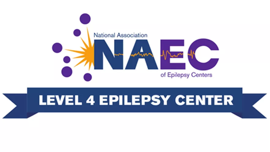
Designación de Centro de Epilepsia de Nivel 4
Somos uno de los primeros centros integrales de epilepsia de nivel 4 del país, la designación de mayor nivel otorgada por la Asociación Nacional de Centros de Epilepsia, que reconoce nuestra capacidad para desempeñarnos en un amplio espectro de subtipos de epilepsia infantil y adulta, y para ofrecer los tratamientos más innovadores y recientes para los casos más complejos. Estamos dentro del 1 % de los mejores hospitales del país en neurología y neurocirugía según U.S. News & World Report.
Nuestro equipo multidisciplinario de epilepsia trata una amplia variedad de afecciones, como astrocitomas, malformaciones cavernosas, displasias, epilepsia, hemangiomas, esclerosis mesial temporal, convulsiones, esclerosis tuberosa, entre muchas otras.
Atención de renombre mundial en todas las etapas de la vida
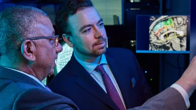
Cada vez se reconoce más que la epilepsia afecta a las interacciones dinámicas entre todo el cerebro y el cuerpo. El Centro Integral de Epilepsia aborda este desafío al tratar a pacientes en todas las etapas de la vida con un equipo líder de epileptólogos, neurocirujanos, neurorradiólogos, neuropsicólogos, neuropatólogos, tecnólogos en electroencefalografía, asesores genéticos y trabajadores sociales de renombre mundial, tanto para pacientes adultos como pediátricos. Esta colaboración integral nos permite profundizar nuestra comprensión de la epilepsia para ofrecer opciones de tratamiento individualizadas en todo el espectro de enfermedades y afecciones del sistema nervioso, desde las más comunes hasta las más complejas.
Tratamiento individualizado avanzado
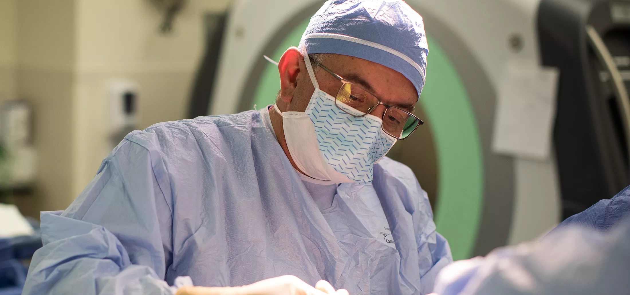
Los tratamientos quirúrgicos para la epilepsia se consideran cuando los medicamentos no logran controlar adecuadamente las convulsiones, afectando de manera significativa la calidad de vida de la persona. No todas las personas con epilepsia son candidatas para una cirugía; esta decisión se basa en una evaluación integral realizada por un equipo de profesionales de la salud compuesto por neurólogos, neurocirujanos, neuropsicólogos y otros especialistas.
Antes de realizar una cirugía para la epilepsia, se lleva a cabo una evaluación exhaustiva que generalmente incluye un monitoreo mediante un videoelectroencefalograma (vEEG), neuroimágenes como una resonancia magnética (MRI, por sus siglas en inglés) y una tomografía por emisión de positrones (TEP, por sus siglas en inglés), pruebas neuropsicológicas, una prueba de Wada (un estudio angiográfico que determina la lateralización del lenguaje y la función de la memoria) y otras pruebas diagnósticas. El objetivo de esta fase inicial es localizar las convulsiones y evaluar la importancia funcional de las áreas afectadas. Estos estudios forman parte de la denominada investigación de fase 1.
Todos los datos recopilados en la fase 1 se analizan durante una Conferencia Multidisciplinaria sobre Epilepsia, donde se establecen recomendaciones consensuadas. En algunos casos, la información obtenida es suficiente para avanzar directamente hacia un tratamiento específico, y en otros casos, se necesita una segunda fase, que implica una investigación más detallada para localizar el foco epiléptico.
A continuación, se detallan algunos de los tratamientos quirúrgicos para la epilepsia que ofrece Montefiore Einstein.
A la vanguardia de la innovación
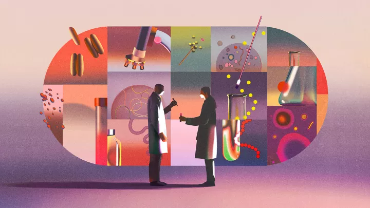
Nuestro equipo de médicos investigadores clínicos ha encabezado ensayos clínicos de epilepsia en niños y adultos a nivel nacional. Por medio de su trabajo colaborativo, han contribuido a comprender los orígenes y la evolución de la epilepsia a lo largo del desarrollo y la vida adulta, así como las diferencias en la manera en que afecta a hombres y mujeres.
También somos líderes en el estudio de las convulsiones febriles, los trastornos del lenguaje y el autismo, así como en la relación entre las convulsiones y las lesiones cerebrales inducidas por estas. Además, estudiamos el tratamiento individualizado de la epilepsia y los trastornos convulsivos, el impacto de las terapias más avanzadas en la calidad de vida y el rendimiento escolar, los espasmos infantiles, la patogénesis y los nuevos tratamientos. Asimismo, exploramos la relación entre el estrés y la epilepsia, así como la antiepileptogénesis o la prevención de la enfermedad.


Su equipo del Centro de Epilepsia


Alexis D. Boro, MD


Nuestros especialistas son reconocidos como líderes nacionales e internacionales en la comunidad de neurocirugía. El doctor Emad Eskandar es presidente de la American Society for Stereotactic and Functional Neurosurgery (ASSFN), el doctor Shlomo Shinnar preside la American Epilepsy Society (AES) y el doctor Solomon Moshe encabeza la Liga Internacional contra la Epilepsia (ILAE, por sus siglas en inglés). El grupo lidera investigaciones pioneras, ha recibido numerosas becas y ha publicado cientos de artículos.
Acerca de la epilepsia
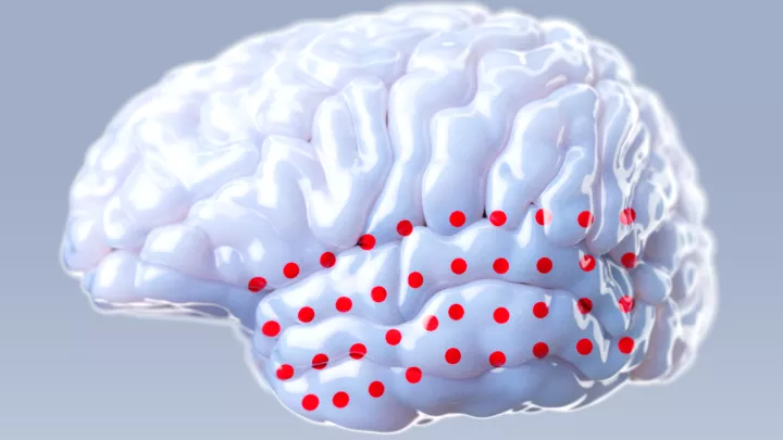
Las epilepsias son trastornos neurológicos crónicos en los que grupos de células nerviosas, o neuronas, del cerebro a veces envían señales anómalas y provocan convulsiones. Las neuronas normalmente generan señales eléctricas y químicas que actúan sobre otras neuronas, glándulas y músculos para producir pensamientos, sentimientos y acciones humanas.
Durante una crisis epiléptica, muchas neuronas y distintas partes del cerebro se sincronizan y emiten señales a la misma frecuencia, lo que significa que temporalmente no pueden realizar sus funciones normales. Este aumento repentino de actividad eléctrica simultánea puede provocar movimientos, sensaciones, emociones o comportamientos involuntarios, y la alteración temporal de la actividad neuronal normal puede causar pérdida de consciencia.
La epilepsia puede considerarse un trastorno del espectro debido a sus diversas causas, los distintos tipos de crisis epilépticas, su variabilidad en gravedad e impacto entre las personas y la diversidad de afecciones coexistentes. Además, existen muchos tipos diferentes de epilepsia, que se originan por diversas causas.