Our Approach to Epilepsy


The Montefiore Einstein Comprehensive Epilepsy Center is internationally recognized for having one of the nation’s best interdisciplinary epilepsy teams and is a national and international referral site for the most complex epilepsy cases. For 40 years, our center has led the world in innovative and interdisciplinary epilepsy care and research through the Departments of Neurology and Neurosurgery, and our world-renowned specialists are thought leaders at the head of all major national and international epilepsy societies, consortiums, and clinical trials.
Epilepsy affects one to two percent of the population and is a major source of disability, psychological problems, and premature deaths. Between 60–70% of patients can achieve seizure control with medications alone, hence medical therapy is the first line of treatment. However, 30–40% of patients don’t respond to medications and continue to have frequent seizures. A considerable amount of data demonstrates that patients who don’t respond to medication should be evaluated for surgery, which has the potential to dramatically reduce or eliminate seizures, and indicate that the best outcomes are achieved when surgery occurs relatively early in the course of the disease, allowing children to develop normally, or adults to continue/resume their education or work. Our approach is to tailor treatment for each patient and to offer the best treatment options, whatever the age, severity, or chronicity of disease.
Level 4 Epilepsy Center Designation
We are one of the first Comprehensive Level 4 Epilepsy Centers in the nation, the highest level designation from the National Association of Epilepsy Centers that recognizes our ability to operate across a broad continuum of diverse child and adult epilepsy subtypes and provide the latest, innovative treatments for the most complex cases. We are ranked in the top 1% of all hospitals in the nation for neurology and neurosurgery, according to U.S. News & World Report.
Our multidisciplinary epilepsy team treats a vast spectrum of conditions including astrocytoma, cavernous malformations, dysplasias, epilepsy, hemangiomas, mesial temporal sclerosis, seizures, tuberous sclerosis, and many others.
World-renowned Care for All Stages of Life
Epilepsy has increasingly been recognized as affecting dynamic interactions between the entire brain and body. The Comprehensive Epilepsy Center meets this challenge by treating patients at all stages of life with a leading team of world-renowned adult and pediatric epileptologists, neurosurgeons, neuroradiologists, neuropsychologists, neuropathologists, EEG technologists, genetic counselors and social workers. This comprehensive collaboration enables us to accelerate our understanding of epilepsy to offer individualized treatment options across the full spectrum of nervous system illnesses and conditions, from the common to the most complex.

Advanced Individualized Treatment
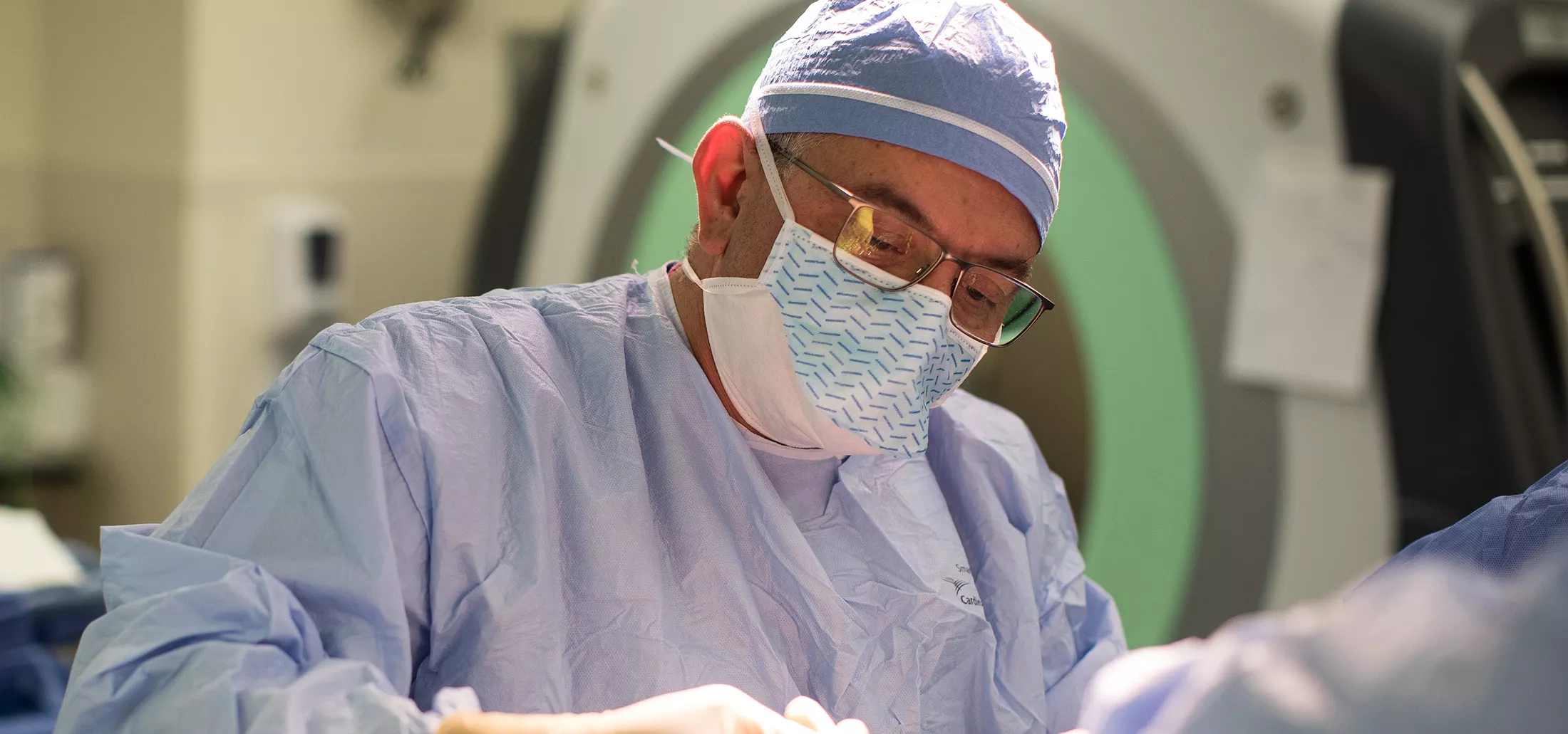
Surgical treatments for epilepsy are considered when medications fail to adequately control seizures, and the seizures significantly impact a person’s quality of life. Not everyone with epilepsy is a candidate for surgery, and the decision to pursue surgical intervention is based on a comprehensive evaluation by a team of healthcare professionals that includes neurologists, neurosurgeons, neuropsychologists, and other specialists.
Before undergoing anterior surgery for epilepsy, a thorough evaluation is conducted. This first phase may include video electroencephalogram (EEG) monitoring, neuroimaging such as magnetic resonance imaging (MRI) and positron emission tomography (PET), neuropsychological testing, a Wada test (an angiographic study that determines lateralization of language and memory function), and other diagnostic tests to pinpoint the location of the seizures and assess the functional significance of the areas to be treated. These studies comprise a Phase-I investigation.
All Phase-I data is reviewed during a Multidisciplinary Epilepsy Conference, where consensus recommendations are made. In some cases, the information is sufficient to proceed directly to a specific treatment, and in other cases, a second phase is required, which involves an investigation to determine the location of the seizure focus.
Following are some of the surgical treatments for epilepsy offered by Montefiore Einstein.
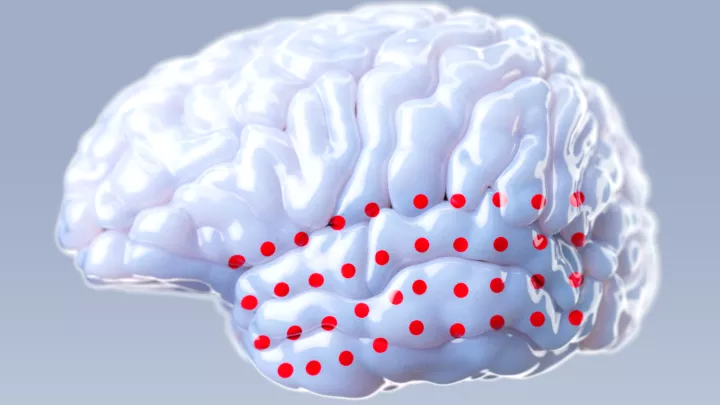
A Phase-II investigation most commonly entails stereoelectroencephalography (SEEG), a neurosurgical technique used for the evaluation and localization of epileptic seizure activity in individuals with drug-resistant epilepsy. SEEG is considered when non-invasive methods, such as scalp EEG (electroencephalogram), fail to provide sufficient information for precise localization of the epileptic focus. SEEG is performed to identify the precise location where seizures originate (the epileptic focus). SEEG allows for the recording of electrical activity deep within the brain without requiring extensive exposure or removal of brain tissue and is considered a minimally invasive procedure.
The SEEG procedure involves the stereotactic (precise) placement of intracranial electrodes directly into the brain to record electrical activity from specific regions. The electrodes used in SEEG are thin, depth electrodes implanted into targeted areas of the brain with robotic assistance. The placement is carefully planned based on preoperative evaluations, including neuroimaging and non-invasive EEG monitoring. Once the electrodes are in place, they continuously record electrical activity from multiple brain regions over an extended period, often for several days. This allows for the capture of spontaneous seizures and the monitoring of brain activity during various tasks and activities.
The device provides detailed information about the epileptic focus and the recorded data from SEEG helps neurologists and neurosurgeons precisely identify the origin of seizures. This information is crucial for planning targeted surgical interventions if surgery is considered a treatment option.
SEEG is typically performed by a multidisciplinary team, including neurologists, neurosurgeons, and epileptologists. The decision to undergo SEEG is based on a careful evaluation of the individual’s specific case and the potential benefits of obtaining detailed information about the epileptic focus for treatment planning. It is an important tool in the comprehensive assessment of drug-resistant epilepsy, particularly when surgical intervention is being considered. Following SEEG, a number of surgical treatments may be considered, including an anterior temporal lobectomy.
An anterior temporal lobectomy (ATL) is one of the most common surgical procedures used to treat epilepsy. It involves the removal of the anterior (front) part of the temporal lobe, typically including the amygdala and hippocampus. ATL is typically considered when seizures are not well-controlled with medications and when they are believed to originate in the medial structures of one temporal lobe.
During the surgery, portions of the anterior part of the anterior temporal lobe, often the hippocampus and amygdala, are removed. The goal is to remove the epileptogenic tissue while preserving as much healthy tissue and function as possible. Often additional recordings are made during the surgery to further refine the surgical procedure.
The success of anterior temporal lobectomy in reducing or eliminating seizures varies among individuals. Between 70–80% of patients experience a significant reduction in seizure frequency or complete seizure freedom, while others may still have some seizures. Individuals considering anterior temporal lobectomy should undergo a comprehensive evaluation by a specialized epilepsy surgery team to determine if they are suitable candidates for the procedure. The decision to undergo surgery should be based on a careful consideration of potential benefits and risks, taking into account the impact of seizures on the individual’s quality of life.
A lesionectomy, or lesion resection, is another possible surgical procedure for patients with epilepsy. This surgery is specifically designed to remove a brain lesion that is believed to be the source of epileptic seizures. It is a possible solution when seizures are associated with a well-defined, localized abnormality in the brain, such as a tumor, vascular malformation, or scar tissue resulting from a previous injury or infection. Lesionectomy may target an abnormality in the frontal, parietal, or temporal lobe.
During the surgery, the lesion is removed while aiming to preserve as much healthy brain tissue as possible. The goal is to eliminate the source of seizures while minimizing the impact on cognitive and neurological function. Intraoperative EEG monitoring may be used to confirm that the removal of the lesion has effectively eliminated abnormal electrical activity in the brain.
The success of a lesionectomy varies among individuals. In cases where the lesion is the primary cause of seizures, removal can lead to a significant reduction in seizure frequency or complete seizure freedom. However, success depends on various factors, including the type and location of the lesion. Individuals considering corpus callosotomy should undergo a comprehensive evaluation by a specialized epilepsy surgery team to determine if they are suitable candidates. The decision to undergo surgery should be based on a careful consideration of potential benefits and risks, taking into account the impact of seizures on the individual’s quality of life.
Another surgical procedure used to treat epilepsy is the corpus callosotomy, which involves the severing or partial cutting of the corpus callosum–the bundle of nerve fibers that connects the two hemispheres of the brain. This procedure is primarily performed as a treatment for certain types of severe epilepsy with uncontrolled generalized seizures, such as atonic (drop) seizures where a sudden loss of muscle tone leads to falls, or tonic-clonic seizures which involve the entire body. This procedure is not commonly performed as a first-line treatment and is usually reserved for cases where other interventions have been ineffective.
Corpus callosotomy is a neurosurgical procedure that is performed under general anesthesia. The surgeon makes an incision in the scalp and drills a small opening in the skull. Using specialized instruments, the corpus callosum is cut or partially severed, disrupting the communication between the two hemispheres of the brain. While corpus callosotomy can reduce the spread of seizures, it does not eliminate the occurrence of seizures themselves. Individuals may still experience seizures, but the impact of the seizures may be less severe. It also does not typically affect consciousness or cognitive function.
The potential benefits of corpus callosotomy include a reduction in the severity and impact of seizures. Individuals considering corpus callosotomy should undergo a comprehensive evaluation by a specialized epilepsy surgery team to determine if they are suitable candidates. The decision to undergo surgery should be based on a careful consideration of potential benefits and risks, taking into account the impact of seizures on the individual’s quality of life.
Some epilepsy patients may qualify for a responsive neurostimulation (RNS), an advanced neurosurgical treatment that involves the implantation of a responsive neurostimulator device to help manage seizures. RNS is designed for individuals with drug-resistant epilepsy who have not achieved sufficient seizure control with medications and are not suitable candidates for resective epilepsy surgery.
During this procedure, two electrodes are stereotactically implanted in or near the seizure onset zone. A small neurostimulator device is surgically implanted in the skull, typically on or near the area of the brain where seizures originate. This neurostimulator continuously monitors the brain’s electrical activity and is programmed to detect abnormal patterns associated with seizures. The device has the ability to differentiate between normal brain activity and the onset of a seizure. When the RNS device detects predefined patterns indicative of an impending seizure, it delivers small electrical pulses or stimulation to the targeted brain area. The goal is to disrupt the abnormal activity and prevent the seizure from fully developing.
The RNS device can be programmed and adjusted by healthcare professionals based on the individual’s specific seizure patterns and response to stimulation. Regular follow-up appointments are typically scheduled to fine-tune the settings and optimize seizure management.
The RNS device continuously logs data related to the patient’s brain activity and seizure events. This information can be downloaded at home or in the office by healthcare providers and assists in the assessment of the effectiveness of the treatment so that informed decisions and adjustments to the therapy can be made.
Responsive neurostimulation is an evolving and promising approach for managing drug-resistant epilepsy. It provides an alternative for individuals who may not be candidates for traditional epilepsy surgery or who have not achieved sufficient seizure control with medications alone. The decision to pursue RNS is individualized and involves a comprehensive assessment by a specialized epilepsy care team.
Some epilepsy patients may benefit from deep brain stimulation (DBS), a neurosurgical technique that involves the implantation of electrodes into specific regions of the brain. This procedure has been used for various neurological and psychiatric conditions and is FDA-approved for the treatment of epilepsy. DBS involves the delivery of electrical impulses to specific brain regions to modulate abnormal neural activity. The goal is to disrupt or prevent the spread of epileptic discharges and reduce the occurrence of seizures. The targets of DBS for epilepsy are in the thalamus, a central nucleus that connects different parts of the brain. Regions targeted for DBS in the context of epilepsy can vary.
Individuals considered for DBS as a treatment for epilepsy are typically those with drug-resistant seizures who have not responded to other treatment options, including medications and surgical interventions. During the procedure, two electrodes are stereotactically implanted, typically in the anterior nucleus of the thalamus onset zone. Two wires are then passed beneath the skin to a small neurostimulator device that is surgically implanted in the chest, below the clavicle.
The effectiveness of DBS can vary among individuals. Some may experience a significant reduction in seizure frequency, while others may have more modest improvements. The benefit increases over several years. As with any surgical procedure, the decision to pursue DBS is based on careful evaluation by a specialized epilepsy team that considers factors such as seizure frequency, seizure type, and the location of the seizure focus.
Vagus nerve stimulation (VNS) is a neuromodulation therapy used in the treatment of epilepsy and is a method that may be considered in particular for individuals with drug-resistant or refractory seizures. The therapy involves the implantation of a device that stimulates the vagus nerve, a major nerve that extends from the brainstem to the abdomen. VNS is designed to help reduce the frequency and severity of seizures.
The VNS procedure involves a small, battery-powered generator implanted under the skin on the chest. A lead wire from the device is wrapped around the left vagus nerve in the neck. The device is programmable and can be adjusted by healthcare professionals to deliver electrical impulses to the vagus nerve at preset intervals. The VNS device stimulates the vagus nerve at regular intervals, typically every few minutes. The electrical impulses travel up the vagus nerve to the brain, where they can help regulate abnormal electrical activity that can lead to seizures.
The device can be programmed to deliver different levels of stimulation based on the individual’s response and needs. Healthcare providers can make adjustments during clinic visits to optimize seizure control while minimizing side effects. Newer VNS devices also have the capability for on-demand or “as- needed” stimulation. In these devices, patients may have a magnet that they can swipe over the generator to initiate extra stimulation if they sense the onset of a seizure or if they are in a situation that may trigger seizures.
The effectiveness of VNS varies. While some people experience a significant reduction in seizure frequency, others may have more modest improvements. It is important to note that VNS does not typically eliminate seizures entirely, but rather aims to reduce their frequency and severity.
VNS is often used as an adjunctive treatment, in addition to an existing antiepileptic medication regimen. It is typically not a standalone treatment and is considered when medications alone are not providing sufficient seizure control. VNS is intended to be a long-term therapy. The decision to pursue VNS is made based on a comprehensive evaluation by a specialized epilepsy care team.

On the Leading Edge of Innovation
Our team of clinical-based physician-researchers has led the nation in clinical trials for pediatric and adult epilepsy, collaborating to understand the origins and changing patterns of epilepsy during development and adult life, as well as the differences between how the disease impacts men and women.
We are also leaders in the study of febrile seizures, language disorders and autism, the relationship between seizures and seizure-induced brain injuries, individualized treatment of epilepsy and seizure disorders, the impact of best treatments on quality of life and school performance, infantile spasms, pathogenesis and new treatments, the relationship between stress and epilepsy, and antiepileptogenesis, or the prevention of epilepsy.

Your Epilepsy Center Team
Our specialists are recognized as national and international leaders in the neurosurgery community. Emad Eskandar, MD, is president of the American Society for Stereotactic and Functional Neurosurgery (ASSFN), Shlomo Shinnar, MD, is president of the American Epilepsy Society (AES), and Solomon Moshe, MD, is president of the International League Against Epilepsy (ILAE). The group is highly involved in research, has been awarded numerous grants and published hundreds of papers.
About Epilepsy
Epilepsies are chronic neurological disorders in which clusters of nerve cells, or neurons, in the brain sometimes signal abnormally and cause seizures. Neurons normally generate electrical and chemical signals that act on other neurons, glands, and muscles to produce human thoughts, feelings, and actions.
During a seizure, many neurons and different parts of the brain become synchronized and fire (signal) at the same frequency, meaning they are temporarily unable to perform their normal functions. This surge of excessive electrical activity happening at the same time may cause involuntary movements, sensations, emotions, or behaviors and the temporary disturbance of normal neuronal activity may cause a loss of awareness.
Epilepsy can be considered a spectrum disorder because of its different causes, different seizure types, its ability to vary in severity and impact from person to person, and its range of coexisting conditions. There also are many different types of epilepsy, resulting from a variety of causes.


