

Video could not be played
Kaskan Laboratory of Amygdala Circuits, Affect & Control Research
Overview
Our work seeks to determine the neural circuits by which stimuli become behaviorally relevant and how that information is represented in the brain, influencing actions and choices and giving rise to human emotional experiences. The human experience also involves learning about “good” and “bad” stimuli and making choices to overcome aversive stimuli and events, which can involve new learning or cognitive mechanisms of control or reappraisal.
Video could not be played
The Role of the Amygdala
The amygdala is a central node in circuits processing affective stimuli that can elicit emotions. Although the amygdala is closely linked to emotional and mental health, it has been difficult to understand its precise role in certain operations and to develop evidence-based interventions in humans because the amygdala is involved in multiple overlapping functions.
These functions likely depend on extensive connections with cortical areas such as the anterior cingulate cortex (ACC), ventromedial prefrontal cortex (vmPFC, sometimes called subgenual ACC in the clinical literature) and orbitofrontal cortex (OFC).
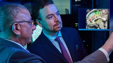
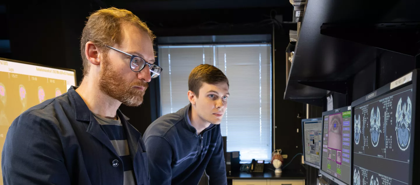
We use the opportunity afforded by human neurosurgical procedures to record local field potentials (LFPs) and single-unit spiking activity from subcortical areas in human patients undergoing deep brain stimulation (DBS) and intracranial recordings (iEEG). Clinically implanted electrodes are used to record data and casually manipulate amygdala and ACC circuits by delivering stimulation during behavioral performance.
Selected Publications
Kaskan PM, Nicholas MA, Dean AM, Murray EA. Attention to stimuli of learned versus innate biological value relies on separate neural systems. J Neurosci. 2022 Dec 7;42(49):9242-9252. doi: 10.1523/JNEUROSCI.0925-22.2022. Epub 2022 Nov 1. PMID: 36319119; PMCID: PMC9761678.
Kaskan PM, Dean AM, Nicholas MA, Mitz AR, Murray EA. Gustatory responses in macaque monkeys revealed with fMRI: Comments on taste, taste preference, and internal state. Neuroimage. 2019 Jan 1;184:932-942. doi: 10.1016/j.neuroimage.2018.10.005. Epub 2018 Oct 3. PMID: 30291973; PMCID: PMC6230501.
Kaskan PM, Costa VD, Eaton HP, Zemskova JA, Mitz AR, Leopold DA, Ungerleider LG, Murray EA. Learned value shapes responses to objects in frontal and ventral stream networks in macaque monkeys. Cereb Cortex. 2017 May 1;27(5):2739-2757. doi: 10.1093/cercor/bhw113. PMID: 27166166; PMCID: PMC6433185.
Baldwin M, Kaskan PM, Zhang B, Chino Y, Kaas JH. Cortical and subcortical connections of V1 and V2 in early postnatal macaque monkeys. J Comp Neurol. 2012 Feb 15;520(3):544-69. doi: 10.1002/cne.22732. PMID: 21800316; PMCID: PMC3673566.
Kaskan PM, Dillenburger BC, Lu HD, Roe AW, Kaas JH. Orientation and direction-of-motion response in the middle temporal visual area (MT) of New World owl monkeys as revealed by intrinsic-signal optical imaging. Front Neuroanat. 2010 Jul 7;4:23. doi: 10.3389/fnana.2010.00023. PMID: 20661299; PMCID: PMC2906256.
Kaskan PM, Lu HD, Dillenburger BC, Kaas JH, Roe AW. The organization of orientation-selective, luminance-change and binocular domains in the second (V2) and third (V3) visual areas of New World owl monkeys as revealed by intrinsic-signal optical imaging. Cereb Cortex. 2009 Jun;19(6):1394-407. doi: 10.1093/cercor/bhn178. Epub 2008 Oct 8. PMID: 18842661; PMCID: PMC2677652.
Kaskan PM, Lu HD, Dillenburger BC, Roe AW, Kaas JH. Intrinsic-signal optical imaging reveals cryptic ocular dominance columns in the primary visual cortex of New World owl monkeys. Front Neurosci. 2007 Oct 15;1(1):67-75. doi: 10.3389/neuro.01.1.1.005.2007. PMID: 18974855; PMCID: PMC2518048.
Kaskan PM, Kaas JH. Cortical connections of the middle temporal and the middle temporal crescent visual areas in prosimian galagos (Otolemur garnetti). Anat Rec (Hoboken). 2007 Mar;290(3):349-66. doi: 10.1002/ar.20440. PMID: 17525950.
Collins CE, Xu X, Khaytin I, Kaskan PM, Casagrande VA, Kaas JH. Optical imaging of visually evoked responses in the middle temporal area after deactivation of primary visual cortex in adult primates. Proc Natl Acad Sci U S A. 2005 Apr 12;102(15):5594-9. doi: 10.1073/pnas.0501762102. Epub 2005 Apr 4. PMID: 15809438; PMCID: PMC556248.
Kaskan PM, Franco EC, Yamada ES, Silveira LC, Darlington RB, Finlay BL. Peripheral variability and central constancy in mammalian visual system evolution. Proc Biol Sci. 2005 Jan 7;272(1558):91-100. doi: 10.1098/rspb.2004.2925. PMID: 15875575; PMCID: PMC1634937.
Xu X, Collins CE, Kaskan PM, Khaytin I, Kaas JH, Casagrande VA. Optical imaging of visually evoked responses in prosimian primates reveals conserved features of the middle temporal visual area. Proc Natl Acad Sci U S A. 2004 Feb 24;101(8):2566-71. doi: 10.1073/pnas.0308745101. PMID: 14983049; PMCID: PMC356990.
Kaskan PM, Finlay BL. Encephalization and its developmental structure: How many ways can a brain get big? In: Falk D, Gibson KR, eds. Evolutionary Anatomy of the Primate Cerebral Cortex. Cambridge: Cambridge University Press; 2001:14-29. doi:10.1017/CBO9780511897085.004.

Video could not be played
Peter M. Kaskan, Ph.D.
About Peter Kaskan, PhD
Dr. Kaskan received his bachelor's degree from Clark University and went on to study at Cornell University and RIKEN Brain Science Institute in Tokyo, Japan. He earned his PhD in psychology (neuroscience) from Vanderbilt University and completed his postdoctoral training at the National Institutes of Health, with partial support from a National Alliance for Research on Schizophrenia & Depression (NARSAD) Young Investigator Grant from the Brain and Behavior Foundation (2014).
Dr. Kaskan joined the faculty of Neurological Surgery in September 2020 after training in intraoperative neurophysiological monitoring for deep brain stimulation at the University of Louisville following his postdoctoral fellowship in the Laboratory of Neuropsychology at the National Institute of Mental Health. Dr. Kaskan received a secondary appointment in Neuroscience at Albert Einstein College of Medicine in August 2023. He collaborates with research groups at the University of Iowa, University of Rochester and Weill-Cornell Medicine.