


Tratamiento de tumores vasculares
En el Centro Oncológico Integral Montefiore Einstein del Hospital Infantil Montefiore Einstein (CHAM) , encontrará atención integral y compasiva para tumores vasculares pediátricos que afectan los vasos sanguíneos y linfáticos. CHAM ofrece atención de vanguardia a niños del área metropolitana de Nueva York, de todo el país y del mundo.
En CHAM, nuestros especialistas en cáncer incluyen médicos de renombre nacional que ofrecen una amplia gama de tratamientos y ensayos clínicos, así como un excepcional equipo de apoyo que forma parte integral de nuestro equipo de atención. Este equipo incluye enfermeros y enfermeras oncológicas especializadas, trabajadores sociales, dietistas registrados, personal de bienestar, fisioterapeutas y terapeutas ocupacionales, especialistas en preservación de la fertilidad (cuando sea apropiado) y especialistas en desarrollo infantil. Nuestro enfoque colaborativo en el tratamiento garantiza que su hijo reciba la mejor atención en un entorno de apoyo y cuidado.
Nuestros esfuerzos en investigación están diseñados para probar nuevas terapias prometedoras, y los ensayos clínicos ofrecen las opciones de tratamiento más avanzadas y actualizadas.
Si lo que usted desea es lo mejor para su hijo, acuda a los especialistas del Centro Oncológico Integral Montefiore Einstein en CHAM, quienes se dedican con pasión a erradicar el cáncer y a atender todas las necesidades de salud de su hijo.

El Montefiore Einstein Comprehensive Cancer Center, designado como centro integral del cáncer por el National Cancer Institute (NCI), apoya la misión y las normas del NCI. La siguiente información sobre los tipos de cáncer, prevención y tratamientos ha sido facilitada por el NCI.
Tumores vasculares infantiles (PDQ®)–Versión para pacientes
¿Qué son los tumores vasculares infantiles?
Los tumores vasculares infantiles son crecimientos anormales de los vasos sanguíneos o linfáticos que pueden aparecer en cualquier parte del cuerpo. Estos tumores pueden ser benignos (es decir, no cancerosos) o cancerosos. Existen muchos tipos de tumores vasculares. El tipo más común es el hemangioma infantil, que es un tumor benigno que generalmente desaparece por sí solo.
Pruebas para diagnosticar tumores vasculares infantiles
Si su hijo o hija presenta síntomas, como decoloración en la piel o debajo de ella, que sugieran la presencia de un tumor vascular, el médico deberá averiguar si se deben a un tumor vascular o a otro problema. El médico le preguntará cuándo comenzaron los síntomas y con qué frecuencia los ha tenido su hijo. También le preguntará sobre los antecedentes de salud personales y familiares de su hijo o hija y le realizará un examen físico . Según estos resultados, es posible que le recomiende otras pruebas. Si a su hijo le diagnostican un tumor vascular, los resultados de estas pruebas le ayudarán a usted y al médico de su hijo a planificar el tratamiento.
Las pruebas utilizadas para diagnosticar un tumor vascular en niños pueden incluir:
Ecografía
Una ecografía utiliza ondas sonoras de alta energía (ultrasonido) que rebotan en los tejidos u órganos internos y producen ecos. Los ecos forman una imagen de los tejidos corporales llamada ecograma .
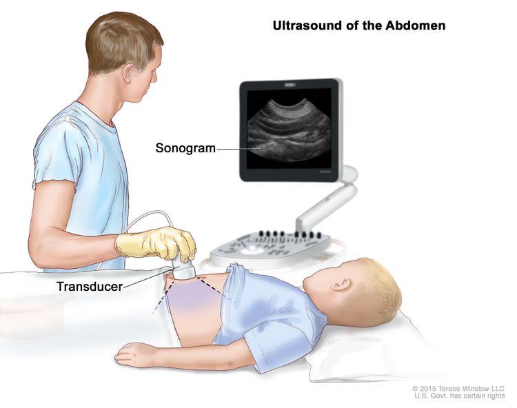
Ultrasonido abdominal: se presiona un transductor de ultrasonido conectado a una computadora contra la piel del abdomen. El transductor hace rebotar ondas de sonido en los órganos y tejidos internos para producir ecos que forman un ecograma (imagen de computadora).
CT scan (CAT scan)
Una tomografía computarizada utiliza una computadora conectada a una máquina de rayos X para crear una serie de imágenes detalladas de áreas internas del cuerpo. Las imágenes se toman desde diferentes ángulos y se utilizan para crear vistas tridimensionales de tejidos y órganos. Se puede inyectar un tinte en una vena o ingerirlo para ayudar a que los órganos o tejidos se vean con más claridad. Este procedimiento también se denomina tomografía computarizada, tomografía computarizada o tomografía axial computarizada. Más información sobre Tomografías computarizadas (TC) y cáncer .
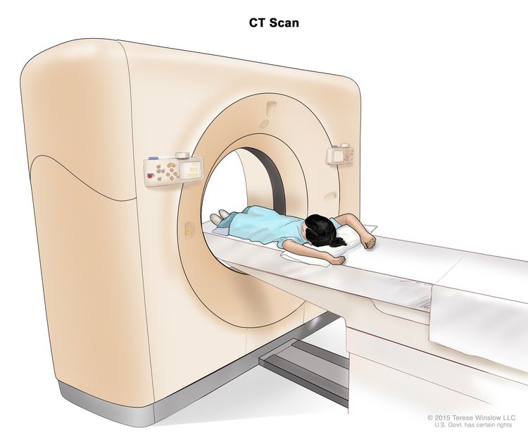
Computed tomography (CT) scan. The child lies on a table that slides through the CT scanner, which takes a series of detailed x-ray pictures of areas inside the body.
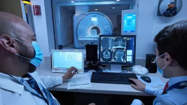
Imágenes por resonancia magnética (IRM)
En la resonancia magnética se utiliza un imán, ondas de radio y una computadora para generar una serie de imágenes detalladas de áreas internas del cuerpo. Este procedimiento también se denomina resonancia magnética nuclear (RMN).
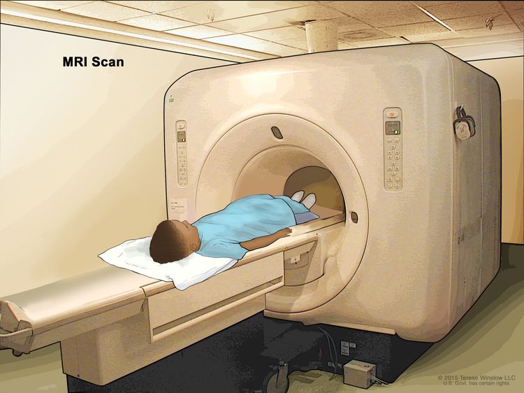
Magnetic resonance imaging (MRI) scan. The child lies on a table that slides into the MRI machine, which takes a series of detailed pictures of areas inside the body. The positioning of the child on the table depends on the part of the body being imaged.
Radiografía de tórax
An x-ray is a type of radiation that can go through the body and make pictures. A chest x-ray makes pictures of the organs and bones inside the chest.
Biopsia
La biopsia es un procedimiento en el que se extrae una muestra de tejido del tumor para que un patólogo pueda observarla bajo un microscopio y detectar si hay cáncer. Si bien no siempre es necesaria una biopsia para diagnosticar un tumor vascular, puede ayudar a encontrar mutaciones genéticas que ayudarán con la planificación del tratamiento.
Pedir una segunda opinión
Puede que quiera pedir una segunda opinión para confirmar el diagnóstico y el plan de tratamiento de su hijo o hija. Si busca una segunda opinión, deberá obtener los resultados de las pruebas médicas y los informes del primer médico para compartirlos con el segundo. El segundo médico revisará el informe patológico, las diapositivas y las exploraciones. Este médico puede estar de acuerdo con el primero, sugerir cambios en el plan de tratamiento o aportar más información sobre el tumor.
Para obtener más información sobre cómo elegir un médico y obtener una segunda opinión, visite Cómo encontrar servicios de atención médica . Puede comunicarse con el Servicio de información sobre el cáncer del NCI a través del chat, correo electrónico o teléfono (tanto en inglés como en español) para obtener ayuda para encontrar un médico u hospital que pueda brindar una segunda opinión. Para preguntas que desee hacer en las citas de su hijo, visite Preguntas para hacerle a su médico .
Tipos de tumores vasculares infantiles
Tumores benignos
Los tumores vasculares benignos no son cáncer.
Hemangioma infantil
El hemangioma infantil (también llamado marca de fresa) es el tipo más común de tumor vascular benigno en los niños. Se produce cuando las células inmaduras que se supone que forman los vasos sanguíneos forman un tumor. Suele aparecer entre las 3 y 6 semanas de edad y, por lo general, no se observa al nacer. El hemangioma suele crecer durante unos 5 meses y luego deja de crecer. Se desvanece lentamente durante los años siguientes, pero puede quedar una marca roja o piel suelta o arrugada. Es poco frecuente que un hemangioma infantil vuelva a aparecer después de que se ha desvanecido.
El hemangioma infantil puede desarrollarse en cualquier parte del cuerpo, incluida la piel, el tejido debajo de la piel o dentro de un órgano. Aparece con mayor frecuencia en la piel de la cabeza y el cuello. El hemangioma puede ser una lesión única, una o más lesiones diseminadas sobre un área más grande o múltiples lesiones en diferentes partes del cuerpo. Un hemangioma que cubre un área más grande, afecta un órgano o tiene múltiples lesiones tiene más probabilidades de causar problemas.
- El hemangioma en las vías respiratorias suele aparecer junto con un hemangioma grande en la cara que parece una barba. El hemangioma de las vías respiratorias puede provocar que estas se estrechen, lo que provoca dificultad para respirar.
- El hemangioma periocular afecta el ojo o los tejidos que lo rodean. Puede causar problemas de visión o ceguera y, a veces, está relacionado con otros problemas oculares.
- La presencia de más de cinco hemangiomas en la piel es un signo de que puede haber hemangiomas en algún órgano, como el hígado, el corazón, los músculos o la glándula tiroides. El hígado es el órgano más afectado.
Algunos hemangiomas aparecen entre las 3 y 6 semanas de vida, pero no crecen. Este tipo de hemangioma se denomina hemangioma infantil con crecimiento mínimo o detenido. La lesión aparece como áreas claras y oscuras de enrojecimiento en la piel de la parte inferior del cuerpo o de la cabeza y el cuello. Los hemangiomas de este tipo desaparecen con el tiempo sin tratamiento.
Causas y factores de riesgo del hemangioma infantil
El hemangioma infantil es causado por ciertos cambios en el funcionamiento de las células vasculares, especialmente en su crecimiento y división en nuevas células. A menudo, se desconoce la causa exacta de estos cambios.
Un factor de riesgo es cualquier cosa que aumenta la probabilidad de contraer una enfermedad. No todos los niños con un factor de riesgo desarrollarán un hemangioma infantil. Y puede desarrollarse en algunos niños que no tienen un factor de riesgo conocido.
El hemangioma infantil es más común en:
- Mujeres jóvenes
- Gente blanca
- Bebés prematuros
- Gemelos, trillizos u otros nacimientos múltiples
- Bebés concebidos mediante fertilización in vitro
- Bebés de madres que son mayores en el momento del embarazo, que tienen preeclampsia ( presión arterial alta durante el embarazo) o que tienen problemas con la placenta durante el embarazo.
Otros factores de riesgo del hemangioma infantil incluyen:
- Tener un historial de salud familiar de hemangioma infantil, generalmente en la madre, el padre o un hermano.
- Padecer el síndrome PHACE, un trastorno poco frecuente que se caracteriza por problemas que afectan los vasos sanguíneos grandes, el corazón, los ojos o el cerebro. El síndrome PHACE aumenta el riesgo de que se desarrolle un hemangioma que se extienda por una zona extensa de la cabeza o la cara y, a veces, por el cuello, el pecho o el brazo.
- Padecer el síndrome LUMBAR/PELVIS/SACRO, un trastorno poco frecuente caracterizado por problemas que afectan el sistema urinario, los genitales, el recto, el ano, el cerebro, la médula espinal y las funciones nerviosas. Este síndrome aumenta el riesgo de que se desarrolle un hemangioma que se extienda a una zona extensa de la espalda baja, los brazos, el pecho o las piernas.
Hable con el médico de su hijo si cree que su hijo puede estar en riesgo.
Síntomas del hemangioma infantil
El hemangioma infantil puede causar cualquiera de los siguientes síntomas. Es importante consultar con el médico de su hijo si su hijo tiene:
- Lesión en la piel: una zona con venas en forma de araña o piel más clara o descolorida puede ser el primer signo de un hemangioma. Esto puede convertirse en una lesión firme, cálida y de color rojo brillante a carmesí en la piel que puede parecer un hematoma. Una lesión también puede formar una úlcera que es dolorosa y puede provocar sangrado, infección y cicatrización. Más tarde, a medida que el hemangioma desaparece, se vuelve más suave y comienza a desvanecerse en el centro antes de aplanarse y perder color.
- Lesión debajo de la piel: una lesión que crece debajo de la piel en la grasa puede verse azul o violeta. Si la lesión es lo suficientemente profunda debajo de la superficie de la piel, es posible que no se vea.
- Lesión en un órgano: Es posible que no haya síntomas si se forma un hemangioma en un órgano.
Estos síntomas pueden deberse a problemas distintos a un hemangioma. La única forma de saberlo es que su hijo consulte a un médico.
Diagnóstico del hemangioma infantil
Generalmente, para diagnosticar un hemangioma infantil basta con un examen físico y los antecedentes médicos personales y familiares. Si el hemangioma tiene un aspecto inusual, se puede realizar una biopsia . Se puede realizar una ecografía si el hemangioma se encuentra en una zona más profunda del cuerpo sin cambios en la piel o si la lesión cubre una zona extensa del cuerpo. A los bebés con cinco o más hemangiomas en la piel se les debe realizar una ecografía del hígado para comprobar si tienen un hemangioma hepático.
Si el hemangioma es parte de un síndrome, se pueden realizar más pruebas, como un ecocardiograma, una resonancia magnética, una angiografía por resonancia magnética y un examen ocular.
Más información sobre estas pruebas en Pruebas para diagnosticar tumores vasculares infantiles .
Tratamiento del hemangioma infantil
La mayoría de los hemangiomas se desvanecen y se reducen sin tratamiento. Si un hemangioma es grande y causa otros problemas de salud, o está en una zona donde podría causar problemas graves si crece, el tratamiento puede incluir lo siguiente:
- Terapia con betabloqueantes, como propranolol, nadolol o atenolol
- Terapia tópica con betabloqueantes para un hemangioma que se encuentra en una zona de la piel
- Terapia con esteroides, que puede utilizarse cuando se inicia la terapia con betabloqueantes o cuando no se pueden utilizar betabloqueantes.
- Cirugía láser, incluida la cirugía con láser de colorante pulsado, que se puede utilizar para un hemangioma que tiene una úlcera o que no ha desaparecido por completo.
- Cirugía para un hemangioma que tiene una úlcera, causa problemas de visión, no ha desaparecido por completo o está en la cara y no ha respondido a otro tratamiento.
- Terapia combinada, como terapia con propranolol y esteroides o terapia con propranolol y betabloqueantes tópicos
Más información sobre estos tratamientos en Tipos de tratamiento para tumores vasculares infantiles .
Hemangioma congénito
El hemangioma congénito es un tumor vascular benigno que comienza a formarse antes del nacimiento y está completamente formado cuando nace el bebé. Generalmente se encuentra en la piel, pero puede estar en otro órgano. Un hemangioma congénito puede presentarse como una erupción de manchas moradas. La piel alrededor de la mancha puede ser más clara.
Existen tres tipos de hemangiomas congénitos. Las diferencias entre los tres tipos se relacionan con la forma en que se encogen (involucionan) con el tiempo:
- El hemangioma congénito de involución rápida (HCIR) desaparece por sí solo entre 12 y 15 meses después del nacimiento. Puede formar una úlcera, sangrar y causar problemas temporales de corazón y coagulación sanguínea . La piel puede verse un poco diferente incluso después de que el hemangioma desaparezca.
- El hemangioma congénito involutivo parcial (HCIP) puede reducirse por sí solo, pero no desaparece por completo.
- El hemangioma congénito no involutivo (HCNI) permanece del mismo tamaño y nunca desaparece por sí solo.
Si su hijo tiene síntomas que sugieren un hemangioma congénito, el médico le preguntará sobre el historial de salud personal de su hijo y le realizará un examen físico y una ecografía para hacer el diagnóstico.
Los tipos de tratamiento que su hijo puede recibir dependen de si el hemangioma congénito se reducirá por sí solo.
- El tratamiento del hemangioma congénito de involución rápida y del hemangioma congénito de involución parcial puede ser la observación.
- El tratamiento del hemangioma congénito no involutivo puede ser una cirugía para extirpar el tumor, dependiendo de dónde se encuentre y de si está causando síntomas.
Más información sobre estos tratamientos en Tipos de tratamiento para tumores vasculares infantiles .
Tumores vasculares benignos del hígado
Los tumores vasculares benignos del hígado pueden ser:
- Una lesión única en una parte del hígado (lesión vascular focal)
- Varias lesiones en una parte del hígado (lesiones hepáticas múltiples)
- Varias lesiones repartidas por diferentes partes del hígado (lesiones hepáticas difusas).
El hígado tiene muchas funciones, entre ellas filtrar la sangre y producir proteínas que ayudan a la coagulación sanguínea . A veces, el tumor puede bloquear o ralentizar el flujo normal de sangre a través del hígado. Cuando esto sucede, la sangre se envía directamente al corazón sin pasar por el hígado. Esta afección se conoce como derivación hepática. Puede causar insuficiencia cardíaca y problemas de coagulación sanguínea.
Si su hijo tiene síntomas que sugieren un tumor vascular benigno del hígado, el médico le preguntará sobre el historial de salud personal de su hijo y le realizará un examen físico y una ecografía para hacer el diagnóstico.
El tratamiento que puede recibir su hijo depende de si tiene una lesión vascular focal, lesiones hepáticas múltiples o lesiones hepáticas difusas.
Una lesión única en una parte del hígado (lesión vascular focal) suele ser un hemangioma congénito que involuciona rápidamente (se encoge) o un hemangioma congénito que no involuciona. Esta lesión se puede diagnosticar antes del nacimiento o poco después del nacimiento del bebé. El tratamiento de este tipo de lesión depende de si se presentan síntomas y puede incluir:
- Observación
- Embolización del hígado para tratar los síntomas.
- Cirugía, para lesiones que no responden a otros tratamientos.
Las lesiones hepáticas múltiples y difusas suelen ser hemangiomas infantiles. Las lesiones hepáticas difusas pueden causar efectos graves, incluidos problemas con las hormonas tiroideas y el corazón. El hígado puede agrandarse, ejercer presión sobre otros órganos y causar más síntomas.
El tratamiento de lesiones hepáticas múltiples puede incluir:
- Observación de lesiones que no causan síntomas.
- Terapia con betabloqueantes ( propranolol ) para lesiones que comienzan a crecer
El tratamiento de las lesiones hepáticas difusas puede incluir:
- Terapia con betabloqueantes (propranolol)
- Quimioterapia
- Terapia con esteroides
- Hepatectomía total y trasplante de hígado, para lesiones que no responden a la terapia farmacológica o para lesiones hepáticas difusas que se están propagando y causando falla orgánica y no hay tiempo para iniciar el tratamiento.
A los niños con lesiones hepáticas difusas se les puede diagnosticar hipotiroidismo causado por un tumor hepático que utiliza más hormona tiroidea. Estos niños pueden necesitar terapia de sustitución de hormona tiroidea.
Más información sobre estos tratamientos en Tipos de tratamiento para tumores vasculares infantiles .
Si una lesión vascular del hígado no responde al tratamiento, se puede realizar una biopsia para ver si el tumor es cáncer.
Hemangioma de células fusiformes
Un hemangioma de células fusiformes contiene células llamadas células fusiformes. Bajo un microscopio, las células fusiformes se ven largas y delgadas. Un hemangioma de células fusiformes es una lesión dolorosa de color marrón rojizo o azulado que suele aparecer en los brazos o las piernas. Puede comenzar como una lesión y convertirse en más lesiones con el paso de los años. Un hemangioma de células fusiformes puede formarse en niños y adultos.
Algunos niños pueden tener un mayor riesgo de desarrollar un hemangioma de células fusiformes. Un factor de riesgo es cualquier cosa que aumente la probabilidad de contraer una enfermedad. No todos los niños con un factor de riesgo desarrollarán un hemangioma de células fusiformes. Y puede desarrollarse en algunos niños que no tienen un factor de riesgo conocido. Los hemangiomas de células fusiformes tienen más probabilidades de desarrollarse en niños con los siguientes síndromes:
- Síndrome de Maffucci, que afecta el cartílago y la piel.
- Síndrome de Klippel-Trenaunay, que afecta los vasos sanguíneos, los tejidos blandos y los huesos.
Hable con el médico de su hijo si cree que su hijo puede estar en riesgo.
Si su hijo presenta síntomas que sugieren un hemangioma de células fusiformes, el médico le preguntará acerca de los antecedentes de salud personales de su hijo y le realizará un examen físico para hacer el diagnóstico. Si es necesario, se pueden realizar pruebas. Más información sobre Pruebas para diagnosticar tumores vasculares infantiles .
Aunque no existe un tratamiento estándar para el hemangioma de células fusiformes, se puede recurrir a la cirugía para extirpar el tumor. El hemangioma de células fusiformes puede volver a aparecer después de la cirugía.
Hemangioma epitelioide
Un hemangioma epitelioide se forma con mayor frecuencia sobre o dentro de la piel, especialmente en la cabeza, pero puede aparecer en otras áreas, como en los huesos. Un hemangioma epitelioide a veces es causado por una lesión. Se presenta en niños y adultos.
En la piel, un hemangioma epitelioide puede aparecer como protuberancias firmes de color rosa a rojo y puede causar picazón. El hemangioma epitelioide del hueso puede causar hinchazón, dolor y debilitamiento del hueso en el área afectada o síntomas de lesión nerviosa. Estos síntomas pueden deberse a problemas distintos a un hemangioma epitelioide. La única forma de saberlo es que su hijo consulte a un médico.
Si su hijo presenta síntomas que sugieren un hemangioma epitelioide, el médico le preguntará acerca de los antecedentes de salud personales de su hijo y le realizará un examen físico para hacer el diagnóstico. También se puede realizar una resonancia magnética, una radiografía o una biopsia. Más información sobre las pruebas para diagnosticar tumores vasculares infantiles .
No existe un tratamiento estándar para el hemangioma epitelioide. El tratamiento puede incluir:
- Cirugía ( legrado o resección )
- Escleroterapia
- Radioterapia en casos raros
El hemangioma epitelioide a menudo reaparece después del tratamiento.
Más información sobre estos tratamientos en Tipos de tratamiento para tumores vasculares infantiles .
Granuloma piógeno
El granuloma piógeno también se denomina hemangioma capilar lobulillar. Es más común en niños mayores y adultos jóvenes, pero puede aparecer a cualquier edad.
El granuloma piógeno a veces es causado por una lesión o por el uso de ciertos medicamentos, incluidas las píldoras anticonceptivas y los retinoides . También puede formarse sin razón conocida dentro de los capilares (los vasos sanguíneos más pequeños), arterias, venas u otros lugares del cuerpo. Algunas lesiones pueden estar asociadas con malformaciones capilares .
El granuloma piógeno es una lesión elevada, de color rojo brillante, que puede ser pequeña o grande y lisa o con bultos. Crece rápidamente durante semanas o meses y puede sangrar mucho. La lesión se encuentra en la superficie de la piel, pero puede formarse en los tejidos debajo de la piel y parecerse a otras lesiones vasculares. Por lo general, solo hay una lesión. A veces, pueden aparecer múltiples lesiones en la misma zona o en diferentes partes del cuerpo. La única forma de saber si estos síntomas son causados por un granuloma piógeno es que su hijo o hija consulte a un médico.
Si su hijo o hija presenta síntomas que sugieren la presencia de un granuloma piógeno, el médico le preguntará acerca de sus antecedentes médicos personales y le realizará un examen físico para hacer el diagnóstico. Si es necesario, se pueden solicitar pruebas. Más información sobre Pruebas para diagnosticar tumores vasculares infantiles .
El granuloma piógeno puede desaparecer sin tratamiento. En ocasiones, es necesario un tratamiento que puede incluir lo siguiente:
- Cirugía ( escisión o curetaje ) para eliminar la lesión.
- Fotocoagulación
- Crioterapia
- Terapia tópica con betabloqueantes (propranolol o timolol)
El granuloma piógeno a menudo reaparece después del tratamiento.
Más información sobre estos tratamientos en Tipos de tratamiento para tumores vasculares infantiles .
Angiofibroma
El angiofibroma es poco frecuente y aparece como protuberancias rojas en la cara. Es una lesión cutánea benigna que suele presentarse con la esclerosis tuberosa, un trastorno hereditario que causa lesiones cutáneas, convulsiones y discapacidades mentales. Hable con el médico de su hijo o hija si cree que su hijo puede tener un angiofibroma.
Si su hijo o hija presenta síntomas que sugieren la presencia de un angiofibroma, el médico le preguntará acerca de su historial médico personal y le realizará un examen físico para hacer el diagnóstico. Si es necesario, se pueden solicitar pruebas. Más información sobre Pruebas para diagnosticar tumores vasculares infantiles .
El tratamiento del angiofibroma puede incluir:
- Cirugía para extirpar el tumor
- Terapia láser
- targeted therapy (Sirolimús)
Más información sobre estos tratamientos en Tipos de tratamiento para tumores vasculares infantiles .
Angiofibroma nasofaríngeo juvenil
El angiofibroma nasofaríngeo juvenil es un tumor que no es cáncer, pero que puede crecer hacia los tejidos cercanos. Es más común en los varones y puede formarse alrededor de la pubertad. El angiofibroma nasofaríngeo juvenil comienza en la cavidad nasal y puede extenderse a la nasofaringe, los senos paranasales, el hueso que rodea los ojos y, a veces, al cerebro.
Si su hijo o hija presenta síntomas que sugieren la presencia de un angiofibroma nasofaríngeo juvenil, el médico le preguntará acerca de los antecedentes de salud personales de su hijo y le realizará un examen físico para llegar al diagnóstico. De ser necesario, se pueden solicitar pruebas. Más información sobre Pruebas para diagnosticar tumores vasculares infantiles .
El tratamiento del angiofibroma nasofaríngeo juvenil puede incluir:
- Cirugía para extirpar el tumor
- Radioterapia
- Quimioterapia
- immunotherapy (Interferón)
- targeted therapy (Sirolimús)
Este tumor puede reaparecer después del tratamiento.
Más información sobre estos tratamientos en Tipos de tratamiento para tumores vasculares infantiles .
Tumores intermedios que pueden propagarse localmente
Es probable que algunos tumores intermedios se propaguen al área alrededor del tumor (localmente), pero no a otras partes del cuerpo.
Hemangioendotelioma kaposiforme y angioma en penacho
El hemangioendotelioma kaposiforme y el angioma en penacho son tumores de los vasos sanguíneos que se presentan en bebés o niños pequeños y afectan a hombres y mujeres por igual. Estos tumores pueden causar el fenómeno de Kasabach-Merritt, una afección en la que la sangre no puede coagularse y puede producirse un sangrado grave. Este tipo de tumor vascular no está relacionado con el sarcoma de Kaposi .
Síntomas del hemangioendotelioma kaposiforme y del angioma en penacho
El hemangioendotelioma kaposiforme y el angioma en penacho suelen aparecer en la piel de los brazos y las piernas, pero también pueden formarse en tejidos más profundos, como músculos o huesos, o en el pecho, el abdomen, la cabeza o el cuello.
Los síntomas pueden ser:
- Áreas de piel firmes, cálidas y dolorosas que parecen amoratadas.
- Zonas de piel de color púrpura o rojo parduzco
- Dolor sin bulto visible
- moretones fáciles
- Sangrado mayor de lo normal en las membranas mucosas, heridas y otros tejidos
Las personas que tienen hemangioendotelioma kaposiforme o angioma en penacho pueden presentar anemia (debilidad, sensación de cansancio o palidez).
Estos síntomas pueden deberse a problemas distintos del hemangioendotelioma kaposiforme o el angioma en penacho. La única forma de saberlo es que su hijo consulte a un médico.
Diagnóstico del hemangioendotelioma kaposiforme y del angioma en penacho
Si un examen físico y una resonancia magnética muestran claramente que el tumor es un hemangioendotelioma kaposiforme o un angioma en penacho, es posible que no sea necesaria una biopsia . No siempre se realiza una biopsia porque puede producirse un sangrado grave. También se puede utilizar una ecografía para diagnosticar un angioma en penacho.
Más información sobre las pruebas para diagnosticar tumores vasculares infantiles .
Tratamiento del hemangioendotelioma kaposiforme y del angioma en penacho
El hemangioendotelioma kaposiforme y el angioma en penacho se tratan mejor con un especialista en anomalías vasculares. El tratamiento depende de los síntomas, el tamaño y la ubicación del tumor, y del riesgo de sangrado. La infección, la demora en el tratamiento y la cirugía pueden causar sangrado potencialmente mortal.
El hemangioendotelioma kaposiforme y el angioma en penacho pueden denominarse no complicados o complicados.
Los tumores sin complicaciones se localizan en una zona, son más pequeños, causan pocos síntomas o ninguno y tienen un menor riesgo de sangrado. Las personas con un tumor sin complicaciones no presentan el fenómeno de Kasabach-Merritt.
El tratamiento del hemangioendotelioma kaposiforme no complicado y del angioma en penacho puede incluir:
- Observación de tumores con bajo riesgo de empeoramiento.
- Cirugía para extirpar el tumor
- laser surgery
- Terapia tópica ( esteroides o tacrolimus )
- Terapia con betabloqueantes ( propranolol )
- Terapia dirigida ( sirolimus ) con o sin terapia con esteroides
Los tumores complicados son más grandes, pueden causar síntomas y afectar el funcionamiento del organismo. Las personas con un tumor complicado pueden padecer el fenómeno de Kasabach-Merritt, una afección grave que puede poner en riesgo la vida y requiere tratamiento.
El tratamiento del hemangioendotelioma kaposiforme complicado y del angioma en penacho puede incluir:
- Quimioterapia, con o sin terapia con esteroides
- Terapia dirigida (sirolimus), con o sin terapia con esteroides
- Cirugía, con o sin embolización
Incluso con tratamiento, estos tumores no desaparecen por completo y pueden volver a aparecer. El dolor y la inflamación pueden empeorar con la edad, a menudo alrededor de la pubertad . Los efectos a largo plazo incluyen dolor crónico, insuficiencia cardíaca, problemas óseos y linfedema (acumulación de líquido linfático en los tejidos).
Más información sobre estos tratamientos en Tipos de tratamiento para tumores vasculares infantiles .
Tumores intermedios que pueden extenderse a otras partes del cuerpo.
En raras ocasiones, los tumores intermedios se propagan a otras partes del cuerpo (hacen metástasis).
Hemangioendotelioma pseudomiogénico (similar al sarcoma epitelioide)
El hemangioendotelioma pseudomiogénico puede presentarse en niños, pero es más común en hombres de 20 a 50 años. Este tumor es poco común y generalmente se presenta sobre o debajo de la piel o en el hueso. El hemangioendotelioma pseudomiogénico puede aparecer como un bulto en el tejido blando o puede causar dolor en el área afectada. Puede propagarse al tejido cercano, pero generalmente no se propaga a otras partes del cuerpo. En la mayoría de los casos, hay múltiples tumores. Hable con el médico de su hijo o hija si cree que su hijo o hija puede tener hemangioendotelioma pseudomiogénico.
Si su hijo o hijapresenta síntomas que sugieren un hemangioendotelioma seudomiogénico, el médico le preguntará acerca de los antecedentes de salud personales de su hijo o hija y le realizará un examen físico para hacer el diagnóstico. De ser necesario, se pueden solicitar pruebas. Más sobre las pruebas para diagnosticar tumores vasculares infantiles .
El tratamiento del hemangioendotelioma pseudomiogénico puede incluir:
- Cirugía para extirpar el tumor cuando sea posible o puede ser necesaria la amputación cuando hay múltiples tumores en el hueso.
- Quimioterapia
- Terapia dirigida (pazopanib)
Debido a que el hemangioendotelioma pseudomiogénico es tan raro en niños, las opciones de tratamiento se basan en ensayos clínicos en adultos.
Más información sobre estos tratamientos en Tipos de tratamiento para tumores vasculares infantiles .
Hemangioendotelioma retiforme
El hemangioendotelioma retiforme es un tumor plano de crecimiento lento que se presenta en adultos jóvenes y, a veces, en niños. Este tumor suele aparecer sobre o debajo de la piel de los brazos, las piernas y el tronco . Por lo general, no se propaga a otras partes del cuerpo. Hable con el médico de su hijo o hija si cree que su hijo puede tener un hemangioendotelioma retiforme.
Si su hijo o hija presenta síntomas que sugieren un hemangioendotelioma retiforme, el médico le preguntará acerca de los antecedentes de salud personales de su hijo o hija y le realizará un examen físico para hacer el diagnóstico. De ser necesario, se pueden solicitar pruebas. Más sobre las pruebas para diagnosticar tumores vasculares infantiles .
El tratamiento del hemangioendotelioma retiforme puede incluir:
- Cirugía para extirpar el tumor
- Radioterapia y quimioterapia cuando no se puede realizar la cirugía o cuando el tumor ha regresado
El hemangioendotelioma retiforme puede reaparecer después del tratamiento.
Más información sobre estos tratamientos en Tipos de tratamiento para tumores vasculares infantiles .
Angioendotelioma papilar intralinfático
El angioendotelioma papilar intralinfático también se denomina tumor de Dabska y se presenta en niños y adultos.
El angioendotelioma papilar intralinfático puede aparecer como protuberancias firmes, elevadas y de color púrpura, que pueden ser pequeñas o grandes. El angioendotelioma papilar intralinfático se forma en la piel o debajo de ella en cualquier parte del cuerpo. A veces, los ganglios linfáticos se ven afectados. Hable con el médico de su hijo o hija si cree que su hijo o hija puede tener angioendotelioma papilar intralinfático.
Si su hijo o hija presenta síntomas que sugieren un angioendotelioma intralinfático papilar, el médico le preguntará acerca de los antecedentes de salud personales de su hijo o hija y le realizará un examen físico para hacer el diagnóstico. De ser necesario, se pueden solicitar pruebas. Más sobre las pruebas para diagnosticar tumores vasculares infantiles .
El tratamiento del angioendotelioma intralinfático papilar es quirúrgico.
Más información sobre este tratamiento en Tipos de tratamiento para tumores vasculares infantiles .
Hemangioendotelioma compuesto
El hemangioendotelioma compuesto tiene características de tumores vasculares tanto benignos como malignos . Este tumor generalmente se presenta en la piel o debajo de ella, en los brazos o las piernas. También puede presentarse en la piel de la cabeza, el cuello o el pecho. Es poco probable que el hemangioendotelioma compuesto se propague a los tejidos cercanos o a otras partes del cuerpo, pero puede reaparecer en el mismo lugar. Si el tumor se disemina, generalmente lo hace a los ganglios linfáticos cercanos. El hemangioendotelioma compuesto se presenta en niños y adultos.
Si su hijo o hija presenta síntomas que sugieren un hemangioendotelioma compuesto, el médico le preguntará acerca de los antecedentes de salud personales de su hijo y le realizará un examen físico para hacer el diagnóstico. Si es necesario, se pueden solicitar pruebas. Más sobre las pruebas para diagnosticar tumores vasculares infantiles .
El tratamiento del hemangioendotelioma compuesto puede incluir:
- Cirugía para extirpar el tumor
- Radioterapia y quimioterapia para tumores que se han diseminado
El hemangioendotelioma compuesto puede reaparecer después del tratamiento.
Más información sobre estos tratamientos en Tipos de tratamiento para tumores vasculares infantiles .
sarcoma de Kaposi
El sarcoma de Kaposi es un cáncer que provoca el crecimiento de lesiones en la piel, las membranas mucosas que recubren la boca, la nariz y la garganta, los ganglios linfáticos u otros órganos. Es causado por el virus del herpes humano 8. Este cáncer rara vez se presenta en niños. En los Estados Unidos, el sarcoma de Kaposi se presenta con mayor frecuencia en niños que tienen un sistema inmunitario débil causado por trastornos poco frecuentes del sistema inmunitario, infección por VIH o medicamentos utilizados en trasplantes de órganos. En África subsahariana, el sarcoma de Kaposi es endémico y a menudo se presenta en niños y adultos jóvenes.
El sarcoma de Kaposi son lesiones que se forman en la piel, la boca o la garganta. Las lesiones son de color rojo, morado o marrón y cambian de ser planas a elevadas, a escamosas, llamadas placas, o a nódulos . A veces, el sarcoma de Kaposi provoca inflamación de los ganglios linfáticos. Estos síntomas pueden deberse a problemas distintos del sarcoma de Kaposi. La única forma de saberlo es consultar al médico de su hijo o hija.
Si su hijo o hija presenta síntomas que sugieren sarcoma de Kaposi, el médico le preguntará acerca de los antecedentes de salud personales de su hijo o hija y le realizará un examen físico para hacer el diagnóstico. Si es necesario, se pueden solicitar pruebas. Más sobre las pruebas para diagnosticar tumores vasculares infantiles .
El tratamiento del sarcoma de Kaposi puede incluir:
Más información sobre estos tratamientos en Tipos de tratamiento para tumores vasculares infantiles .
Debido a que el sarcoma de Kaposi es tan inusual en niños, algunas opciones de tratamiento se basan en ensayos clínicos en adultos. Obtenga más información en Tratamiento del sarcoma de Kaposi .
Tumores malignos
Los tumores malignos son cáncer.
Epithelioid hemangioendothelioma
El hemangioendotelioma epitelioide puede presentarse en niños, pero es más común en adultos de 30 a 50 años. Puede presentarse en el hígado, los pulmones, los huesos, la piel o los tejidos blandos. El hemangioendotelioma epitelioide puede crecer rápido o lentamente. En aproximadamente un tercio de los pacientes con un tumor en los tejidos blandos, el tumor se propaga a otras partes del cuerpo muy rápidamente.
Síntomas del hemangioendotelioma epitelioide
Los síntomas del hemangioendotelioma epitelioide dependen de la ubicación del tumor en el cuerpo. Es importante consultar con el médico de su hijo o hija si presenta:
- Manchas de color marrón rojizo en la piel que están elevadas y redondeadas o son planas y se sienten calientes.
- Síntomas tempranos de lesiones en el pulmón, que pueden no presentarse en todos los niños con lesiones pulmonares:
- chest pain
- Escupir sangre
- Anemia (debilidad, sensación de cansancio o palidez)
- Dificultad para respirar (debido a tejido pulmonar cicatrizado)
- Huesos rotos
- Síntomas de lesiones en el hígado:
- Picor
- Coloración amarillenta de la piel o los ojos
Estos síntomas pueden deberse a problemas distintos a un hemangioendotelioma epitelioide. La única forma de saberlo es que su hijo o hija consulte a un médico.
Diagnóstico del hemangioendotelioma epitelioide
Si su hijo o hija tiene síntomas que sugieren la presencia de un hemangioendotelioma epitelioide, el médico le preguntará acerca de los antecedentes de salud personales de su hijo y le realizará un examen físico para hacer el diagnóstico. El médico también puede solicitar pruebas. El hemangioendotelioma epitelioide en el hígado se detecta mediante una ecografía, una tomografía computarizada o una resonancia magnética . También pueden realizarse radiografías del tórax o de otras áreas del cuerpo. Más información sobre Pruebas para diagnosticar tumores vasculares infantiles .
Tratamiento del hemangioendotelioma epitelioide
El tratamiento del hemangioendotelioma epitelioide de crecimiento lento puede ser la observación. Se puede recurrir a la cirugía cuando sea posible extirpar el tumor.
El tratamiento del hemangioendotelioma epitelioide de crecimiento rápido puede incluir:
- Cirugía para extirpar el tumor cuando sea posible.
- Inmunoterapia ( interferón ) para tumores que tienen probabilidades de propagarse
- Terapia dirigida ( pazopanib o sirolimus ) para tumores que tienen probabilidades de propagarse
- Quimioterapia
- Radioterapia
- palliative care
Más información sobre estos tratamientos en Tipos de tratamiento para tumores vasculares infantiles .
Angiosarcoma
El angiosarcoma es un tumor de rápido crecimiento que se forma en los vasos sanguíneos o linfáticos de cualquier parte del cuerpo, generalmente en el tejido blando. La mayoría de los angiosarcomas se encuentran en la piel o en el tejido blando cercano a la piel. Los que se encuentran en el tejido blando más profundo pueden formarse en el hígado, el bazo y el pulmón.
El angiosarcoma es muy poco frecuente en niños. A veces, los niños tienen más de un tumor en la piel, el hígado o ambos.
Causas y factores de riesgo del angiosarcoma
Un factor de riesgo es cualquier cosa que aumenta la probabilidad de contraer una enfermedad. No todos los niños con un factor de riesgo desarrollarán angiosarcoma. Y puede desarrollarse en algunos niños que no tienen un factor de riesgo conocido. Los factores de riesgo del angiosarcoma incluyen:
- being exposed to radiation
- Linfedema crónico (a largo plazo), una afección en la que se acumula líquido linfático adicional en los tejidos y causa hinchazón
- Tener un tumor vascular benigno
En raras ocasiones, un tumor vascular benigno, como un hemangioma, puede convertirse en un angiosarcoma.
Hable con el médico de su hijo si cree que su hijo puede estar en riesgo.
Síntomas del angiosarcoma
Los síntomas del angiosarcoma dependen de dónde se encuentre el tumor y pueden incluir:
- Manchas rojas en la piel que sangran fácilmente.
- Tumores morados
Estos síntomas pueden deberse a problemas distintos a un angiosarcoma. La única forma de saberlo es que su hijo consulte a un médico.
Diagnóstico del angiosarcoma
Para hacer un diagnóstico, el médico le preguntará acerca de los antecedentes de salud personales de su hijo y le realizará un examen físico. Si es necesario, se pueden solicitar pruebas. Más sobre las pruebas para diagnosticar tumores vasculares infantiles .
Tratamiento del angiosarcoma
El tratamiento del angiosarcoma puede incluir:
- surgery to completely remove the tumor
- Una combinación de cirugía, quimioterapia y radioterapia para el angiosarcoma que se ha diseminado
- palliative care
Más información sobre estos tratamientos en Tipos de tratamiento para tumores vasculares infantiles .
Tipos de tratamiento para los tumores vasculares infantiles
¿Quién trata a los niños con tumores vasculares?
Un oncólogo pediátria, médico especializado en el tratamiento de niños con cáncer, supervisa el tratamiento de tumores vasculares infantiles. El oncólogo pediátrico colabora con otros profesionales de la salud expertos en el tratamiento de niños con cáncer y que también se especializan en ciertas áreas de la medicina. Otros especialistas pudieran incluir:
- Especialista en anomalías vasculares pediátricas (experto en el tratamiento de niños con tumores vasculares)
- pediatric surgeon
- Dermatólogo pediatra
- Hematólogo y oncólogo pediátra
- Cirujano ortopédico
- Oncólogo radioterapeuta
- Especialista en enfermería pediátrica
- Especialista en rehabilitación
- Psicólogo
- Trabajador social
Opciones de tratamiento
Existen diferentes tipos de tratamiento para niños y adolescentes con tumores vasculares. Usted y el equipo médico de su hijo colaborarán para decidir el tratamiento. Se considerarán diversos factores, como la ubicación del tumor, la edad y el estado de salud general de su hijo, el tipo de tumor vascular, el riesgo de cicatrización y la probabilidad de tratamiento completo del tumor vascular.
El plan de tratamiento de su hijo o hija incluirá información sobre el cáncer, los objetivos terapéuticos, las opciones disponibles y los posibles efectos secundarios. Es recomendable hablar con el equipo médico antes de que comience el tratamiento para conocer qué esperar en cada etapa. Para obtener ayuda en cada paso del proceso, consulte nuestro folleto descargable Niños con cáncer: una guía para padres.
Los tipos de tratamiento que su hijo o hija podría recibir son:
Terapia con betabloqueantes
Los betabloqueantes son medicamentos que se usan comúnmente para reducir la presión arterial y la frecuencia cardíaca, pero también pueden reducir el tamaño de ciertos tipos de tumores vasculares, como los hemangiomas infantiles. La terapia con betabloqueantes puede inyectarse en una vena, administrarse por vía oral o aplicarse sobre la piel ( tópica ). La forma de administrar la terapia con betabloqueantes depende del tipo de tumor vascular que se esté tratando y de dónde se formó inicialmente.
El betabloqueante propranolol suele ser el primer tratamiento para los hemangiomas. Los bebés menores de 4 semanas que presentan una afección subyacente o que reciben tratamiento con propranolol intravenoso podrían necesitar iniciar el tratamiento en un hospital. El hemangioma infantil también puede tratarse con propranolol y terapia con esteroides, o con propranolol y betabloqueantes tópicos. El propranolol también se utiliza para tratar tumores vasculares benignos del hígado.
Otros betabloqueantes utilizados para tratar tumores vasculares incluyen atenolol, nadolol y timolol.
Cirugía
Los siguientes tipos de cirugía se pueden utilizar para extirpar muchos tipos de tumores vasculares:
- La escisión es una cirugía para extirpar todo el tumor y parte del tejido sano que lo rodea.
- La cirugía láser utiliza un haz de luz (un haz estrecho de luz intensa) como bisturí para realizar cortes sin sangrado en el tejido o extirpar una lesión cutánea, como un tumor. La cirugía con láser de colorante pulsado puede utilizarse para algunos hemangiomas. Este tipo de láser utiliza un haz de luz que se dirige a los vasos sanguíneos de la piel. La luz se transforma en calor y los vasos sanguíneos se destruyen sin dañar la piel circundante.
- El curetaje utiliza un instrumento pequeño en forma de cuchara con un borde afilado llamado cureta para eliminar tejido anormal.
- La hepatectomía total y el trasplante de hígado extraen todo el hígado seguido de un trasplante de un hígado sano de un donante .
- La amputación elimina un brazo o una pierna cuando hay múltiples tumores en el hueso.
El tipo de cirugía utilizada depende del tipo de tumor vascular y de dónde se formó en el cuerpo.
Después de que el médico extirpe todo el cáncer visible en el momento de la cirugía, algunas personas podrían recibir quimioterapia o radioterapia para eliminar cualquier célula cancerosa restante. El tratamiento que se administra después de la cirugía para reducir el riesgo de reaparición del cáncer se denomina terapia adyuvante .
Fotocoagulación
La fotocoagulación consiste en el uso de un haz de luz intenso, como un láser, para sellar vasos sanguíneos o destruir tejido. Se utiliza para tratar el granuloma piógeno .
Cryotherapy
La crioterapia utiliza un instrumento para congelar y destruir tejido anormal, como los vasos sanguíneos anormales en el granuloma piógeno. Este tipo de tratamiento también se denomina criocirugía.
Learn more about Cryosurgery to Treat Cancer.
Embolización
La embolización utiliza partículas, como pequeñas esponjas o microesferas de gelatina, para obstruir los vasos sanguíneos del hígado. Puede utilizarse para bloquear el flujo sanguíneo a algunos tumores vasculares benignos del hígado y al hemangioendotelioma kaposiforme.
Quimioterapia
La quimioterapia (también llamada quimio) utiliza medicamentos para detener el crecimiento de las células tumorales. La quimioterapia destruye las células cancerosas o impide que se multipliquen. La quimioterapia puede administrarse sola o en combinación con otros tipos de tratamiento.
Para algunos tumores vasculares, la quimioterapia se inyecta en una vena. Al administrarse de esta manera, los medicamentos ingresan al torrente sanguíneo y llegan a las células tumorales de todo el cuerpo.
Los medicamentos de quimioterapia que pueden usarse solos o en combinación para tratar tumores vasculares infantiles incluyen:
También pueden utilizarse otros medicamentos quimioterápicos no incluidos en esta lista.
Para obtener más información sobre el efecto, la forma de administración, los efectos secundarios comunes y otros datos de la quimioterapia, consulte Quimioterapia para tratar el cáncer.
Sclerotherapy
La escleroterapia destruye el tumor y los vasos sanguíneos que lo forman. Se inyecta un líquido en los vasos sanguíneos, lo que provoca su cicatrización y destrucción. Con el tiempo, los vasos sanguíneos destruidos son absorbidos por el tejido sano. La sangre fluye por las venas sanas cercanas. La escleroterapia se utiliza para tratar el hemangioma epitelioide.
Radioterapia
La radioterapia utiliza rayos X de alta energía u otros tipos de radiación para destruir las células tumorales o impedir su crecimiento. La radioterapia externa utiliza una máquina externa al cuerpo para enviar radiación hacia la zona del cuerpo donde se encuentra el tumor. Se utiliza para tratar algunos tumores vasculares.
Para obtener más información, consulte Radioterapia de haz externo para el cáncer y Efectos secundarios de la radioterapia.
Terapia dirigida
La terapia dirigida utiliza medicamentos u otras sustancias para bloquear la acción de enzimas, proteínas u otras moléculas específicas que participan en el crecimiento y la propagación de las células cancerosas. Se utilizan o se estudian diferentes tipos de terapia dirigida para tratar los tumores vasculares infantiles:
Obtenga más información en Terapia dirigida para tratar el cáncer.
Inmunoterapia
La inmunoterapia ayuda al sistema inmunitario a combatir el cáncer. El interferón es un tipo de inmunoterapia que se utiliza para tratar tumores vasculares.
Obtenga más información sobre la inmunoterapia para el tratamiento del cáncer .
Other drug therapy
Otros medicamentos utilizados para tratar tumores vasculares infantiles o controlar sus efectos incluyen:
- Terapia con esteroides: Los esteroides son hormonas que el cuerpo produce de forma natural. También pueden producirse en un laboratorio y usarse como medicamentos. Los esteroides ayudan a reducir el tamaño de algunos tumores vasculares. Los corticosteroides, como la prednisona y la metilprednisolona, se utilizan para tratar el hemangioma infantil.
- Terapia inmunosupresora: Estos medicamentos reducen la respuesta inmunitaria del organismo. La terapia inmunosupresora se ha utilizado para reducir el tamaño de los tumores vasculares. El tacrolimus tópico se utiliza para tratar los hemangioendoteliomas kaposiformes y los angiomas en penacho.
- Terapia de sustitución de hormona tiroidea: estos medicamentos reemplazan las hormonas producidas por la tiroides y se utilizan para tratar una forma rara de hipotiroidismo causada por algunos tumores vasculares, como los hemangiomas hepáticos.
Observación
La observación consiste en monitorear de cerca la condición de una persona sin administrar ningún tratamiento hasta que los síntomas aparezcan o se modifiquen.
Ensayos clínicos
Para algunos niños, participar en un ensayo clínico puede ser una opción. Hay distintos tipos de ensayos clínicos para el cáncer infantil. Por ejemplo, un ensayo de tratamiento prueba nuevos tratamientos o nuevas formas de utilizar los tratamientos actuales. Los ensayos de atención de apoyo y cuidados paliativos buscan formas de mejorar la calidad de vida, especialmente para quienes presentan efectos secundarios del cáncer y su tratamiento.
Puede usar la búsqueda de ensayos clínicos para encontrar ensayos clínicos sobre cáncer financiados por el NCI que aceptan participantes. Esta búsqueda le permite filtrar los ensayos según el tipo de cáncer, la edad de su hijo o hija y el lugar donde se realizan. Puede encontrar ensayos clínicos financiados por otras organizaciones en el sitio web ClinicalTrials.gov.
Para más información sobre ensayos clínicos, cómo encontrarlos y participar en uno de ellos, visite la web Información sobre estudios clínicos para pacientes y cuidadores.
Efectos secundarios y efectos tardíos del tratamiento
Los tratamientos para tumores vasculares pueden causar efectos secundarios. Los efectos secundarios que su hijo pueda experimentar dependerán del tipo de tratamiento que reciba, la dosis y la reacción de su cuerpo. Hable con el equipo de tratamiento de su hijo sobre los efectos secundarios a los que debe prestar atención y cómo controlarlos.
Para obtener más información sobre los efectos secundarios que aparecen durante el tratamiento del cáncer, consulte la sección Efectos secundarios.
Los problemas derivados del tratamiento del cáncer que comienzan 6 meses o más tarde y continúan durante meses o años se denominan efectos tardíos. Estos efectos pueden incluir:
- Problemas físicos
- Cambios en el estado de ánimo, los sentimientos, los pensamientos, el aprendizaje o la memoria
- segundos cánceres (nuevos tipos de cáncer)
Algunos efectos tardíos pueden tratarse o controlarse. Es importante hablar con los médicos de su hijo sobre los posibles efectos tardíos causados por algunos tratamientos. Obtenga más información sobre los efectos tardíos del tratamiento del cáncer infantil .
Cuidados de seguimiento
A medida que su hijo/a continúa con el tratamiento, se le realizarán pruebas o chequeos de seguimiento. Es posible que se repitan algunas de las pruebas realizadas para diagnosticar el tumor vascular para evaluar la eficacia del tratamiento. Las decisiones sobre si continuar, modificar o suspender el tratamiento pueden basarse en los resultados de estas pruebas.
Algunas pruebas se seguirán realizando periódicamente después de finalizar el tratamiento. Los resultados de estas pruebas pueden indicar si la condición de su hijo ha cambiado o si el tumor ha reaparecido.
Más información sobre las pruebas de seguimiento en Pruebas para diagnosticar tumores vasculares infantiles .
Cómo afrontar el tumor vascular de su hijo
Cuando su hijo tiene un tumor, todos los miembros de la familia necesitan apoyo. Cuidarse durante este momento difícil es importante. Busque apoyo en el equipo de tratamiento de su hijo, así como en familiares y comunidades. Para obtener más información, visite Apoyo para Familias: Cáncer Infantil y el folleto Niños con Cáncer: Una Guía para Padres .
Related resources
Para obtener más información sobre el cáncer infantil y otros recursos generales sobre el cáncer, consulte los siguientes sitios web:
Sobre este resumen del PDQ
Acerca del PDQ
El Physician Data Query (PDQ) es la base de datos integral sobre el cáncer del National Cancer Institute (NCI). La base de datos del PDQ contiene resúmenes con la última información publicada sobre prevención, detección, genética, tratamiento, atención médica de apoyo y medicina complementaria y alternativa relacionada con el cáncer. La mayoría de los resúmenes se presentan en dos versiones. Las versiones para profesionales de la salud contienen información detallada escrita en lenguaje técnico. Las versiones para pacientes están escritas en un lenguaje fácil de entender y no tan técnico. Ambas versiones contienen información precisa y actualizada sobre el cáncer. La mayoría de las versiones también están disponibles en español.
El PDQ es un servicio del NCI. El NCI es parte de los Institutos Nacionales de Salud (NIH), que son el centro de investigación biomédica del Gobierno federal. Los resúmenes del PDQ se basan en una revisión independiente de la literatura médica. No son declaraciones de políticas del NCI ni de los NIH.
Propósito de este resumen
Este resumen de información sobre el cáncer del PDQ contiene información actualizada sobre el tratamiento de tumores vasculares infantiles. Su objetivo es informar y ayudar a los pacientes, las familias y los cuidadores. No proporciona directrices ni recomendaciones formales para tomar decisiones sobre la atención médica.
Revisores y actualizaciones
Los comités editoriales escriben los resúmenes de información sobre el cáncer del PDQ y los mantienen actualizados. Estos comités están formados por equipos de especialistas en el tratamiento del cáncer y otras especialidades relacionadas con esta enfermedad. Los resúmenes se revisan periódicamente y se modifican cuando hay información nueva. La fecha de actualización al pie de cada resumen indica cuándo se realizó el cambio más reciente.
La información de este resumen para pacientes procede de la versión para profesionales de la salud, la cual es revisada y actualizada periódicamente por el comité editorial del PDQ sobre el tratamiento pediátrico según sea necesario.
Información sobre ensayos clínicos
Un ensayo clínico es un estudio para responder a una pregunta científica como, por ejemplo, si un tratamiento es mejor que otro. Los ensayos se basan en estudios anteriores y en lo aprendido en el laboratorio. Cada ensayo responde a determinadas preguntas científicas que permiten encontrar nuevas y mejores formas de ayudar a los pacientes con cáncer. Durante los ensayos clínicos de tratamiento, se recopila información sobre los efectos de un nuevo tratamiento y su eficacia. Si un ensayo clínico demuestra que un nuevo tratamiento es mejor que uno que se utiliza actualmente, el nuevo tratamiento puede convertirse en “estándar”. Los pacientes pueden valorar la posibilidad de participar en un ensayo clínico. Algunos ensayos clínicos solo están abiertos a pacientes que no hayan iniciado el tratamiento.
Los ensayos clínicos se pueden encontrar en línea en el sitio web del NCI. Para obtener más información, llame al Servicio de Información sobre el Cáncer (CIS, por sus siglas en inglés), el centro de contacto del NCI, al 1-800-4-CANCER (1-800-422-6237).
Permiso de uso de este resumen
Physician Data Query (PDQ) es una marca registrada. Se autoriza el libre uso del contenido de los documentos del PDQ como texto. Sin embargo, no se podrá identificar como un resumen de información sobre cáncer del PDQ del NCI, salvo que se reproduzca en su totalidad y se actualice con regularidad. Por otra parte, se permite que los autores incluyan una oración como “en el resumen del PDQ del NCI sobre la prevención del cáncer de mama se describen, de manera concisa, los siguientes riesgos: [incluir fragmento del resumen]”.
La forma recomendada para citar este resumen del PDQ es:
Comité editorial del PDQ® sobre el tratamiento pediátrico. Tumores vasculares infantiles del PDQ. Bethesda, MD: National Cancer Institute. Actualizado el [DD/MM/AAAA].
Las imágenes de este resumen se utilizan con el permiso del autor, artista y/o editorial para uso exclusivo en los resúmenes del PDQ. Si desea usar una imagen de un resumen del PDQ sin incluir el resumen completo, debe obtener autorización del propietario. El National Cancer Institute no puede otorgar dicho permiso. Para obtener más información sobre el uso de las imágenes de este resumen o de otras ilustraciones relacionadas con el cáncer, consulte Visuals Online, una colección de más de 3,000 imágenes científicas.
Descargo de responsabilidad
La información de estos resúmenes no debe utilizarse para tomar decisiones sobre reembolsos de seguros. Puede encontrar más información sobre la cobertura de seguros en Cancer.gov en el sitio Manejo de la atención del cáncer.
Contáctenos
Puede encontrar más información sobre cómo contactarnos o recibir ayuda en el sitio web Cancer.gov en la página Comuníquese con el NCI. También puede enviar sus preguntas a Cancer.gov en el apartado Escríbanos del sitio web.
Actualizado:
URL de origen: https://www.cancer.gov/node/1050998/syndication
Agencia de origen: National Cancer Institute (NCI)
Fecha de captura: 2016-06-03 07:39:47.0