


Tumores del sistema nervioso central
En el Centro Oncológico Integral Montefiore Einstein, encontrará atención médica excepcional para tumores del sistema nervioso central que afectan el cerebro o la médula espinal. Como uno de los primeros centros oncológicos designados por el NCI, durante más de 50 años hemos sido líderes en la investigación, el diagnóstico y el tratamiento de más de 200 tipos de cáncer.
Encuentre atención integral para tumores del sistema nervioso central en el equipo especializado de Neurooncología del Centro Oncológico Integral Montefiore Einstein. Nuestros reconocidos especialistas en cáncer de cerebro y médula espinal lideran investigaciones de vanguardia y trabajan en conjunto para garantizar que su plan de tratamiento se adapte a sus necesidades. Apreciará nuestro enfoque holístico y centrado en el paciente, así como los recursos de apoyo que le ayudarán a mantener la mejor calidad de vida y alcanzar su máximo potencial de salud.
Si necesita atención médica, confíe en nuestros profesionales, que se dedican con pasión a acabar con el cáncer y a atender todas sus necesidades de salud.

El Montefiore Einstein Comprehensive Cancer Center, designado como centro integral del cáncer por el National Cancer Institute (NCI), apoya la misión y las normas del NCI. La siguiente información sobre los tipos de cáncer, prevención y tratamientos ha sido facilitada por el NCI.
Tratamiento de los tumores del sistema nervioso central en adultos (PDQ®): versión para pacientes
Información general sobre los tumores del sistema nervioso central en adultos
Puntos clave
- Un tumor del sistema nervioso central (SNC) en adultos es una enfermedad en la que se forman células anómalas en los tejidos del encéfalo y/o la médula espinal.
- Un tumor que comienza en otra parte del cuerpo y se disemina al encéfalo se llama tumor cerebral metastásico.
- El encéfalo controla muchas funciones corporales importantes.
- La médula espinal conecta el encéfalo con los nervios en la mayor parte del cuerpo.
- Hay diferentes tipos de tumores cerebrales y de la médula espinal.
- Tumores astrocíticos
- Tumores oligodendrogliales
- Gliomas mixtos
- Tumores ependimarios
- Meduloblastomas
- Tumores del parénquima pineal
- Tumores meníngeos
- Tumores de células germinales
- Craneofaringioma (grado 1)
- Tener ciertos síndromes genéticos puede aumentar el riesgo de desarrollar un tumor del SNC.
- Se desconoce la causa de la mayoría de los tumores del encéfalo y de la médula espinal en adultos.
- Los signos y síntomas de los tumores del encéfalo y de la médula espinal en adultos no son los mismos en todas las personas.
- Las pruebas que examinan el encéfalo y la médula espinal se utilizan para diagnosticar tumores cerebrales y de la médula espinal en adultos.
- También se puede realizar una biopsia para diagnosticar un tumor cerebral.
- A veces no es posible realizar una biopsia o una cirugía.
- Certain factors affect prognosis (chance of recovery) and treatment options.
Un tumor del sistema nervioso central (SNC) en adultos es una enfermedad en la que se forman células anómalas en los tejidos del encéfalo y/o la médula espinal.
Existen muchos tipos de tumores cerebrales y de médula espinal . Estos tumores se forman por el crecimiento anormal de células y pueden originarse en diferentes partes del cerebro o la médula espinal. El cerebro y la médula espinal, en conjunto, conforman el sistema nervioso central (SNC).
Los tumores pueden ser benignos (no cancerosos) o malignos (cancerosos):
- Los tumores cerebrales y de la médula espinal benignos crecen y presionan áreas cercanas del encéfalo. No suelen propagarse a otros tejidos y pueden recidivar (regresar).
- Es probable que los tumores cerebrales y de la médula espinal malignos crezcan rápidamente y se propaguen a otros tejidos del encéfalo.
Cuando un tumor crece o presiona un área del cerebro, puede impedir que esa parte del cerebro funcione correctamente. Tanto los tumores cerebrales benignos como los malignos producen signos y síntomas y necesitan tratamiento.
Los tumores cerebrales y de la médula espinal pueden desarrollarse tanto en adultos como en niños. Sin embargo, el tratamiento para niños puede ser diferente al que se usa en adultos.
Para obtener información sobre el linfoma que comienza en el cerebro, consulte Tratamiento del linfoma primario del SNC .
Un tumor que comienza en otra parte del cuerpo y se disemina al encéfalo se llama tumor cerebral metastásico.
Los tumores que comienzan en el cerebro se denominan tumores cerebrales primarios. Los tumores cerebrales primarios pueden diseminarse a otras partes del cerebro o a la columna vertebral. No suelen propagarse a otras partes del cuerpo.
Con frecuencia, los tumores cerebrales se originan en otra parte del cuerpo y se diseminan a una o más áreas del cerebro. Estos se denominan tumores cerebrales metastásicos (o metástasis cerebrales). Los tumores cerebrales metastásicos son más comunes que los tumores cerebrales primarios. Hasta la mitad de los tumores cerebrales metastásicos provienen de cáncer de pulmón .
El cáncer puede diseminarse a las leptomeninges (las dos membranas más internas que cubren el cerebro y la médula espinal). Esto se denomina carcinomatosis leptomeníngea.
El encéfalo controla muchas funciones corporales importantes.
El encéfalo tiene tres partes principales:
- El cerebro es la parte más grande del encéfalo. Está en la parte superior de la cabeza. El cerebro controla el pensamiento, el aprendizaje, la resolución de problemas, las emociones, el habla, la lectura, la escritura y el movimiento voluntario.
- El cerebelo se encuentra en la parte inferior posterior del encéfalo (cerca de la mitad de la parte posterior de la cabeza). Controla el movimiento, el equilibrio y la postura.
- El tronco del encéfalo conecta el encéfalo con la médula espinal. Está en la parte más baja del encéfalo (justo encima de la parte posterior del cuello). El tronco del encéfalo controla la respiración, la frecuencia cardiaca y los nervios y músculos que se utilizan para ver, oír, caminar, hablar y comer.
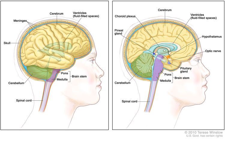
Anatomía del encéfalo que muestra el cerebro, los ventrículos (con el líquido cefalorraquídeo en azul), el cerebelo, el tronco encefálico (puente y bulbo raquídeo) y otras partes del encéfalo.
La médula espinal conecta el encéfalo con los nervios en la mayor parte del cuerpo.
La médula espinal es una columna de tejido nervioso que va desde el tronco encefálico hasta el centro de la espalda. Está cubierto por tres finas capas de tejido llamadas membranas, las cuales están rodeadas por las vértebras (huesos de la espalda). Los nervios de la médula espinal transportan mensajes entre el encéfalo y el resto del cuerpo, como una señal enviada por el cerebro para hacer que los músculos se muevan o de la piel al cerebro para percibir el tacto.
Hay diferentes tipos de tumores cerebrales y de la médula espinal.
Los tumores cerebrales y de la médula espinal reciben nombres según el tipo de célula en la que se formaron y el lugar en el que se formó el tumor por primera vez en el SNC. El grado de un tumor se puede utilizar para diferenciar entre los de crecimiento lento y de crecimiento rápido. Los grados de tumores de la Organización Mundial de la Salud (OMS) se basan en cuán anómalas se ven las células cancerosas bajo un microscopio y en la rapidez con la que es probable que el tumor crezca y se propague.
Sistema de gradación de tumores de la OMS
- Grado 1 (grado bajo): las células tumorales se parecen más a las células normales bajo un microscopio y crecen y se propagan más lentamente que las células tumorales de grado 2, 3 y 4. Rara vez se propagan a los tejidos cercanos. Los tumores cerebrales de grado 1 se pueden extirpar completamente mediante cirugía.
- Grado 2: las células tumorales crecen y se diseminan más lentamente que las células tumorales de grado 3 y 4. Pueden diseminarse al tejido cercano y recidivar (regresar). Algunos tumores pueden convertirse en tumores de mayor grado.
- Grado 3: las células tumorales se ven muy diferentes de las células normales bajo el microscopio y crecen más rápidamente que las células tumorales de grado 1 y 2. Es probable que se propaguen al tejido cercano.
- Grado 4 (alto grado): las células tumorales no parecen células normales bajo un microscopio y crecen y se diseminan muy rápidamente. Puede haber zonas de células muertas en el tumor. Los tumores de grado 4 generalmente no se pueden extirpar completamente mediante cirugía.
Los siguientes tipos de tumores primarios se pueden formar en el encéfalo o la médula espinal:
Tumores astrocíticos
Un tumor astrocítico se origina en las células cerebrales con forma de estrella, llamadas astrocitos, que ayudan a mantener a las neuronas sanas. Un astrocito es un tipo de célula glial. Las células gliales a veces forman tumores llamados gliomas. Los tumores astrocíticos pueden ser:
- Glioma de tronco encefálico (generalmente de alto grado): un glioma de tronco encefálico se forma en el tronco encefálico, que es la parte del encéfalo conectada a la médula espinal. A menudo es un tumor de alto grado, que afecta extensamente el tronco encefálico. Los gliomas del tronco encefálico son raros en adultos.
- Tumor astrocítico pineal (cualquier grado): un tumor astrocítico pineal se forma en el tejido alrededor de la glándula pineal y puede ser de cualquier grado. La glándula pineal es un pequeño órgano del cerebro que produce melatonina, una hormona que ayuda a controlar el ciclo de sueño y vigilia.
- Astrocitoma pilocítico (grado 1): un astrocitoma pilocítico crece lentamente en el cerebro o la médula espinal. Puede tener la forma de un quiste y rara vez se propaga a los tejidos cercanos.
- Astrocitoma difuso (grado 2): un astrocitoma difuso crece lentamente, pero a menudo se disemina a los tejidos cercanos. Las células tumorales se parecen a las células normales. También recibe el nombre de astrocitoma difuso de bajo grado.
- Astrocitoma anaplásico (grado 3): un astrocitoma anaplásico crece rápidamente y se propaga a los tejidos circundantes. Las células tumorales tienen un aspecto diferente al de las células normales. También se denomina astrocitoma maligno o astrocitoma de alto grado.
- Glioblastoma (grado 4): un glioblastoma crece y se propaga muy rápidamente. Las células tumorales se ven muy diferentes de las células normales. También recibe el nombre de glioblastoma multiforme.
Tumores oligodendrogliales
Un tumor oligodendroglial se origina en las células cerebrales llamadas oligodendrocitos, que ayudan a mantener a las neuronas sanas. Un oligodendrocito es un tipo de célula glial. Los oligodendrocitos a veces forman tumores llamados oligodendrogliomas. Los grados de tumores oligodendrogliales son:
- Oligodendroglioma (grado 2): un oligodendroglioma crece lentamente, pero a menudo se propaga a los tejidos cercanos. Las células tumorales se parecen a las células normales.
- Oligodendroglioma anaplásico (grado 3): un oligodendroglioma anaplásico crece rápidamente y se propaga a los tejidos cercanos. Las células tumorales se ven diferentes de las células normales.
Gliomas mixtos
Un glioma mixto es un tumor cerebral que tiene dos tipos de células tumorales: oligodendrocitos y astrocitos. Este tipo de tumor mixto se llama oligoastrocitoma.
- Oligoastrocitoma (grado 2): un oligoastrocitoma es un tumor de crecimiento lento. Las células tumorales se parecen a las células normales.
- Oligoastrocitoma anaplásico (grado 3): un oligoastrocitoma anaplásico crece rápidamente y se disemina a los tejidos cercanos. Las células tumorales tienen un aspecto diferente al de las células normales. Este tipo de tumor tiene un pronóstico menos favorable que el oligoastrocitoma (grado 2).
Tumores ependimarios
Un tumor ependimario suele originarse en las células que recubren los espacios llenos de líquido del cerebro y alrededor de la médula espinal. Un tumor ependimario también se denomina ependimoma. Los grados de ependimomas son:
- Ependimoma (grado 1 o 2): un ependimoma de grado 1 o 2 crece lentamente y tiene células que se parecen a las células normales. Hay dos tipos de ependimoma de grado 1: ependimoma mixopapilar y subependimoma. Un ependimoma de grado 2 crece en un ventrículo (espacio lleno de líquido en el cerebro) y sus vías de conexión o en la médula espinal.
- Ependimoma anaplásico (grado 3): un ependimoma anaplásico crece rápidamente y se propaga a los tejidos cercanos. Las células tumorales se ven diferentes de las células normales. Este tipo de tumor suele tener peor pronóstico que un ependimoma de grado 1 o 2.
Meduloblastomas
El meduloblastoma es un tipo de tumor embrionario. Los meduloblastomas son más comunes en niños o adultos jóvenes.
Para obtener más información sobre los meduloblastomas en niños, consulte el sitio web Tratamiento del meduloblastoma infantil y otros tumores embrionarios del sistema nervioso central.
Tumores del parénquima pineal
Un tumor parenquimatoso pineal se forma en las células parenquimatosas o pineocitos, que son las células que constituyen la mayor parte de la glándula pineal. Estos tumores son diferentes de los tumores astrocíticos pineales. Los grados de tumores del parénquima pineal son:
- Pineocitoma (grado 2): un pineocitoma es un tumor pineal de crecimiento lento.
- Pineoblastoma (grado 4): un pineoblastoma es un tumor poco común que tiene probabilidades altas de diseminarse.
Para obtener más información sobre los tumores del parénquima pineal en niños, consulte el sitio web Tratamiento del meduloblastoma infantil y otros tumores embrionarios del sistema nervioso central.
Tumores meníngeos
Un tumor meníngeo, también llamado meningioma, se forma en las meninges (capas delgadas de tejido que cubren el cerebro y la médula espinal). Puede formarse a partir de diferentes tipos de células del cerebro o de la médula espinal. Los meningiomas son más comunes en adultos. Los tipos de tumores meníngeos son:
- Meningioma (grado 1): un meningioma de grado 1 es el tipo más común de tumor meníngeo. Es un tumor de crecimiento lento. Se forma con mayor frecuencia en la duramadre. Un meningioma de grado 1 se puede extirpar completamente mediante cirugía.
- Meningioma (grado 2 y 3): Este es un tumor meníngeo raro. Crece rápidamente y es probable que se propague dentro del cerebro y la médula espinal. El pronóstico es peor que el de un meningioma de grado 1 porque, por lo general, el tumor no se puede extirpar por completo mediante cirugía.
Un hemangiopericitoma no es un tumor meníngeo pero se trata como un meningioma de grado 2 o 3. Generalmente se forma en la duramadre. El pronóstico es menos favorable que el de un meningioma de grado 1 porque, por lo general, el tumor no se puede extirpar por completo mediante cirugía.
Tumores de células germinales
Un tumor de células germinales se forma en las células germinales, que son las células que se desarrollan en espermatozoides en los hombres u óvulos en las mujeres. Existen diferentes tipos de tumores de células germinales, entre los que se incluyen los germinomas, los teratomas, los carcinomas del saco vitelino embrionario y los coriocarcinomas . Los tumores de células germinales pueden ser benignos o malignos.
Para obtener más información sobre los tumores de células germinales en el cerebro infantil, consulte Tratamiento de los tumores de células germinales del sistema nervioso central infantil.
Craneofaringioma (grado 1)
Un craneofaringioma es un tumor poco común que generalmente se forma en el centro del cerebro, justo encima de la glándula pituitaria (un órgano del tamaño de un guisante en la parte inferior del cerebro que controla otras glándulas). Los craneofaringiomas se pueden formar a partir de diferentes tipos de células del cerebro o de la médula espinal.
Para obtener más información sobre el craneofaringioma en niños, consulte Tratamiento del craneofaringioma infantil.
Tener ciertos síndromes genéticos puede aumentar el riesgo de desarrollar un tumor del SNC.
Cualquier factor que aumente la probabilidad de contraer una enfermedad se denomina factor de riesgo . No todas las personas con uno o más de estos factores de riesgo desarrollarán un tumor cerebral o de médula espinal, y estos pueden desarrollarse en personas sin factores de riesgo conocidos. Consulte con su médico si cree que podría estar en riesgo. Existen pocos factores de riesgo conocidos para los tumores cerebrales. Las siguientes afecciones pueden aumentar el riesgo de ciertos tipos de tumores cerebrales:
- La exposición al cloruro de vinilo puede aumentar el riesgo de glioma.
- La infección por el virus de Epstein-Barr, el SIDA o el trasplante de órganos pueden aumentar el riesgo de linfoma primario del SNC . Para obtener más información, consulte Tratamiento del linfoma primario del SNC .
- Tener ciertos afecciones genéticas síndromes puede aumentar el riesgo de desarrollar tumores cerebrales:
- Neurofibromatosis tipo 1 (NF1) o 2 (NF2).
- enfermedad de von Hippel-Lindau.
- Esclerosis tuberosa.
- Síndrome de Li-Fraumeni.
- Síndrome de Turcot tipo 1 o 2
- Síndrome de carcinoma basocelular nevoide.
Se desconoce la causa de la mayoría de los tumores del encéfalo y de la médula espinal en adultos.
Los signos y síntomas de los tumores del encéfalo y de la médula espinal en adultos no son los mismos en todas las personas.
Signs and symptoms depend on the following:
- Where the tumor forms in the brain or spinal cord.
- Lo que controla la parte del encéfalo afectada.
- El tamaño del tumor
Estos y otros signos y síntomas pueden ser causados por tumores del sistema nervioso central o por otras afecciones, incluido el cáncer que se ha propagado al cerebro. Consulte con su médico si presenta alguno de los siguientes síntomas:
Síntomas de los tumores cerebrales
- Dolor de cabeza matinal o dolor de cabeza que desaparece después de vomitar
- Convulsiones.
- Vision, hearing, and speech problems.
- Pérdida de apetito
- Náuseas y vómitos frecuentes
- Cambios en la personalidad, el estado de ánimo, la capacidad de concentración o el comportamiento
- Pérdida del equilibrio y dificultad para caminar
- Debilidad
- Somnolencia inusual o cambio en el nivel de actividad
Síntomas de los tumores de la médula espinal
- Dolor de espalda o un dolor que se extiende desde la espalda hacia los brazos o las piernas
- Un cambio en los hábitos intestinales o dificultad para orinar
- Debilidad o entumecimiento en los brazos o piernas
- Problemas para caminar
Las pruebas que examinan el encéfalo y la médula espinal se utilizan para diagnosticar tumores cerebrales y de la médula espinal en adultos.
In addition to asking about your personal and family health history and doing a physical exam, your doctor may perform the following tests and procedures:
- Examen neurológico: consiste en una serie de preguntas y pruebas para verificar el funcionamiento del cerebro, la médula espinal y la función nerviosa. El examen analiza el estado mental, la coordinación y la capacidad de una persona para caminar normalmente, así como qué tan bien funcionan los músculos, los sentidos y los reflejos. Esto también puede denominarse examen neurológico.
- Examen del campo visual: es un examen para evaluar el campo visual (el área total en la que se pueden ver los objetos). Esta prueba mide tanto la visión central (cuánto puede ver una persona al mirar al frente) como la visión periférica (cuánto puede ver en todas las demás direcciones al mirar al frente). Cualquier pérdida de visión puede ser señal de un tumor que ha dañado o presionado las partes del cerebro que afectan la vista.
- Prueba de marcadores tumorales: es un procedimiento en el que se toma una muestra de sangre, orina o tejido para medir la cantidad de ciertas sustancias producidas por órganos, tejidos o células tumorales del cuerpo. Ciertas sustancias se relacionan con tipos específicos de cáncer cuando se detectan en niveles elevados en el cuerpo. Estas se denominan marcadores tumorales. Esta prueba puede realizarse para diagnosticar un tumor de células germinales.
- Prueba genética: es una prueba de laboratorio en la que se analizan células o tejidos para detectar cambios en genes o cromosomas. Estos cambios pueden indicar que una persona tiene o está en riesgo de desarrollar una enfermedad o afección específica.
- CT scan (CAT scan): procedimiento que toma una serie de fotografías detalladas de zonas internas del cuerpo desde diferentes ángulos. Las imágenes son tomadas por una computadora conectada a una máquina de rayos X. Un dye may be inyecta en una vena o ingerirlo para permitir que los órganos o tejidos se visualicen con más claridad. Este procedimiento también se llama tomografía axial computarizada.
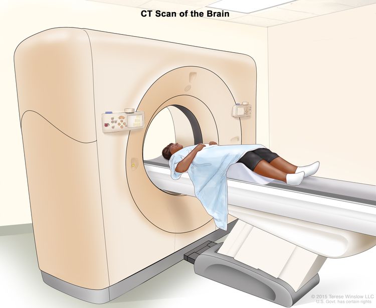
Tomografía computarizada (TC) del cerebro. El paciente se acuesta en una mesa que se desliza a través del escáner CT, que toma imágenes de rayos X del cerebro.
- Imágenes por resonancia magnética (IRM) con gadolinio: es un procedimiento que utiliza un imán, ondas de radio y una computadora para generar una serie de imágenes detalladas del cerebro y la médula espinal. Se inyecta una sustancia llamada gadolinio por una vena. El gadolinio se acumula alrededor de las células cancerosas para que se vean más brillantes en la imagen. Este procedimiento también se llama resonancia magnética nuclear (NMRI). La resonancia magnética se utiliza a menudo para diagnosticar tumores en la médula espinal. A veces, durante la exploración por resonancia magnética se realiza un procedimiento llamado espectroscopia de resonancia magnética (MRS). Una MRS se utiliza para diagnosticar tumores en función de su composición química.
- Exploración SPECT (tomografía computarizada por emisión de fotón único): procedimiento para encontrar células tumorales malignas en el cerebro. Se inyecta una pequeña cantidad de una sustancia radiactiva en una vena o se inhala por la nariz. A medida que la sustancia viaja por la sangre, una cámara gira alrededor de la cabeza y toma fotografías del cerebro. Una computadora usa las imágenes para crear una imagen tridimensional (3D) del cerebro. Habrá un mayor flujo sanguíneo y más actividad en las áreas donde crecen las células cancerosas. Estas áreas aparecerán más brillantes en la imagen.
- Tomografía por emisión de positrones (PET): es un procedimiento que se utiliza para detectar células tumorales malignas en el cuerpo. Se inyecta una pequeña cantidad de glucosa (azúcar) radiactiva en una vena. El escáner PET gira alrededor del cuerpo y crea una imagen de dónde se usa la glucosa en el cerebro. Las células tumorales malignas aparecen más brillantes en la imagen porque son más activas y absorben más glucosa que las células normales. La exploración PET se usa para diferenciar entre un tumor primario y un tumor que se diseminó al cerebro desde otra parte del cuerpo.
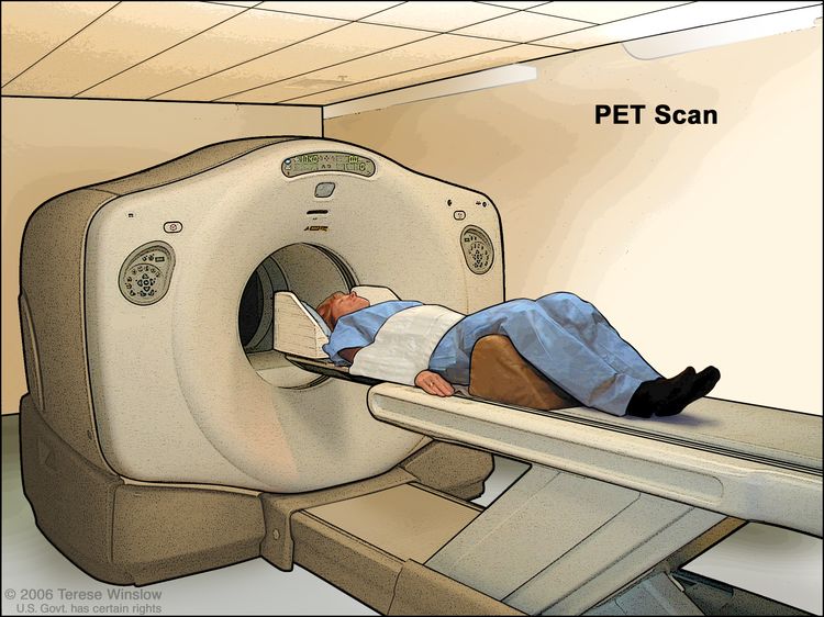
Tomografía por emisión de positrones (PET, por sus siglas en inglés). El paciente se acuesta en una mesa que se desliza a través de la máquina PET. El reposacabezas y la correa blanca ayudan al paciente a permanecer quieto. Se inyecta una pequeña cantidad de glucosa radiactiva (azúcar) en la vena del paciente y un escáner genera una imagen de dónde se usa la glucosa en el cuerpo. Las células cancerosas aparecen más brillantes en la imagen porque absorben más glucosa que las células sanas.
También se puede realizar una biopsia para diagnosticar un tumor cerebral.
Si las pruebas de imagen muestran que puede haber un tumor cerebral, generalmente se realiza una biopsia. Se puede utilizar uno de los siguientes tipos de biopsias:
- Biopsia estereotáctica: Cuando las pruebas de imagen muestran la posible presencia de un tumor en una zona profunda del cerebro de difícil acceso, se puede realizar una biopsia cerebral estereotáctica. Este tipo de biopsia utiliza un ordenador y un escáner tridimensional (3D) para localizar el tumor y guiar la aguja que se utiliza para extraer el tejido. Se realiza una pequeña incisión en el cuero cabelludo y se perfora un pequeño orificio en el cráneo . A través de este orificio, se inserta una aguja de biopsia para extraer células o tejidos que un patólogo podrá examinar al microscopio para detectar signos de cáncer.
- Biopsia abierta: cuando las pruebas de diagnóstico por imágenes muestran que podría haber un tumor que se puede extirpar mediante cirugía, se puede realizar una biopsia abierta. Se extirpa una parte del cráneo en una operación llamada craneotomía. Se extrae una muestra de tejido cerebral y un patólogo la observa al microscopio. Si detecta células cancerosas, es posible que se extirpe una parte o la totalidad del tumor durante la misma cirugía. Se realizan pruebas antes de la cirugía para encontrar las áreas alrededor del tumor que son importantes para el funcionamiento normal del cerebro. También hay formas de evaluar la función cerebral durante la cirugía. El médico usará los resultados de estas pruebas para extirpar la mayor cantidad de tumor posible con el menor daño al tejido normal del cerebro.
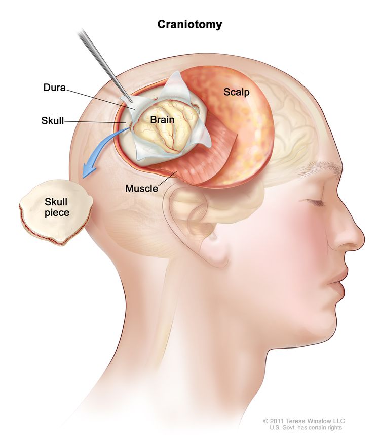
Craneotomía. Se realiza una abertura y se extrae una pieza del cráneo para exponer una parte del cerebro.
El patólogo revisa la muestra de la biopsia para determinar el tipo y grado del tumor cerebral. El grado del tumor se basa en el aspecto de las células tumorales bajo el microscopio y en la rapidez con la que es probable que el tumor crezca y se propague.
Se pueden realizar las siguientes pruebas en el tejido tumoral extirpado:
- Inmunohistoquímica: es una prueba de laboratorio que utiliza anticuerpos para detectar ciertos antígenos (marcadores) en una muestra de tejido de un paciente. Los anticuerpos suelen estar unidos a una enzima o un tinte fluorescente. Después de que los anticuerpos se unen a un antígeno específico en la muestra de tejido, la enzima o el tinte se activa y el antígeno se puede observar bajo un microscopio. Este tipo de prueba se usa para diagnosticar el cáncer y ayudar a distinguir el tipo de cáncer.
- Microscopía óptica y electrónica: prueba de laboratorio en la que se observan las células de una muestra de tejido bajo microscopios regulares y de alta potencia para buscar ciertos cambios en las células.
- Análisis citogenético: prueba de laboratorio en la que se cuentan los cromosomas de las células de una muestra de tejido cerebral y se comprueba si hay cambios, como cromosomas fragmentados, faltantes, reorganizados o adicionales. Los cambios en ciertos cromosomas pueden ser un signo de cáncer. El análisis citogenético se utiliza para ayudar a diagnosticar el cáncer, planificar el tratamiento o determinar su eficacia.
A veces no es posible realizar una biopsia o una cirugía.
En algunos tumores, no se puede realizar una biopsia o una cirugía de manera segura debido al lugar del cerebro o de la médula espinal en el que se formó el tumor. Estos tumores se diagnostican y tratan según los resultados de las pruebas de imagen y otros procedimientos.
A veces, los resultados de las pruebas de imágenes y otros procedimientos muestran que es muy probable que el tumor sea benigno y no se realiza una biopsia.
Certain factors affect prognosis (chance of recovery) and treatment options.
El pronóstico y las opciones de tratamiento para los tumores primarios del cerebro y de la médula espinal dependen de lo siguiente:
- El tipo y grado del tumor
- Dónde está el tumor en el cerebro o la médula espinal.
- Si el tumor se puede extirpar mediante cirugía.
- Whether cancer cells remain after surgery.
- Si hay ciertos cambios en los cromosomas.
- Si el cáncer se acaba de diagnosticar o recidivó (volvió).
- La salud general del paciente
El pronóstico y las opciones de tratamiento para los tumores de encéfalo y de médula espinal metastásicos dependen de lo siguiente:
- Si hay más de dos tumores en el cerebro o la médula espinal.
- Dónde está el tumor en el cerebro o la médula espinal.
- Qué tan bien responde el tumor al tratamiento.
- Si el tumor primario sigue creciendo o propagándose.
Estadios de los tumores del sistema nervioso central en adultos
Puntos clave
- No existe un sistema de estadificación estándar para los tumores cerebrales y de la médula espinal en adultos.
- Las pruebas de imagen pueden repetirse después de la cirugía para ayudar a planificar más tratamientos.
- Los tumores del sistema nervioso central (SNC) suelen recidivar, a veces muchos años después del tratamiento.
No existe un sistema de estadificación estándar para los tumores cerebrales y de la médula espinal en adultos.
El proceso que se utiliza para determinar si el cáncer se ha diseminado a otras áreas del cerebro o a otras partes del cuerpo se denomina estadificación . Los tumores cerebrales que se originan en el cerebro rara vez se diseminan a otras partes del cuerpo. No existe un sistema de estadificación estándar para los tumores cerebrales y de la médula espinal .
El tratamiento de los tumores primarios del cerebro y de la médula espinal se basa en lo siguiente:
- El tipo de célula en la que comenzó el tumor.
- Donde se formó el tumor en el cerebro o la médula espinal.
- La cantidad de cáncer que queda después de la cirugía.
- El grado del tumor
El tratamiento de los tumores que se han diseminado al cerebro desde otras partes del cuerpo se basa en la cantidad de tumores que hay en el cerebro.
Las pruebas de imagen pueden repetirse después de la cirugía para ayudar a planificar más tratamientos.
Algunas de las pruebas y procedimientos utilizados para diagnosticar un tumor cerebral o de la médula espinal pueden repetirse después del tratamiento para determinar cuánto tumor queda.
Los tumores del sistema nervioso central (SNC) suelen recidivar, a veces muchos años después del tratamiento.
Un tumor recurrente del SNC es un tumor que ha reaparecido (regresado) después de haber sido tratado. El tumor puede recividar en el mismo lugar que el tumor original o en otras partes del SNC.
Treatment Option Overview
Puntos clave
- Existen diferentes tipos de tratamiento para pacientes con tumores del cerebro y de la médula espinal en adultos.
- The following types of treatment are used:
- Vigilancia activa
- Cirugía
- Radioterapia
- Quimioterapia
- Terapia dirigida
- Se ofrece atención de apoyo para aliviar los problemas causados por la enfermedad o su tratamiento.
- New types of treatment are being tested in clinical trials.
- Radioterapia con haz de protones
- Inmunoterapia
- El tratamiento de los tumores del sistema nervioso central en adultos puede producir efectos secundarios.
- Patients may want to think about taking part in a clinical trial.
- Patients can enter clinical trials before, during, or after starting their cancer treatment.
- Pueden ser necesarias pruebas de seguimiento.
Existen diferentes tipos de tratamiento para pacientes con tumores del cerebro y de la médula espinal en adultos.
Existen diferentes tipos de tratamiento para pacientes adultos con tumores cerebrales y de médula espinal . Algunos tratamientos son estándar (el tratamiento que se utiliza actualmente) y otros se están probando en ensayos clínicos . Un ensayo clínico es un estudio de investigación cuyo objetivo es mejorar los tratamientos actuales u obtener información sobre nuevos tratamientos para pacientes con cáncer. Cuando los ensayos clínicos demuestran que un nuevo tratamiento es mejor que el estándar, este puede convertirse en el tratamiento estándar. Los pacientes pueden considerar participar en un ensayo clínico. Algunos ensayos clínicos están abiertos únicamente a pacientes que aún no han comenzado el tratamiento.
The following types of treatment are used:
Vigilancia activa
La vigilancia activa consiste en observar atentamente el estado del paciente, pero sin administrar ningún tratamiento a menos que los resultados de las pruebas muestren un empeoramiento de su condición. La vigilancia activa puede utilizarse para evitar o retrasar la necesidad de tratamientos como la radioterapia o la cirugía, que pueden causar efectos secundarios u otros problemas. Durante la vigilancia activa, se realizan ciertos exámenes y pruebas periódicamente. La vigilancia activa puede utilizarse para tumores de crecimiento muy lento que no producen síntomas .
Cirugía
La cirugía puede utilizarse para diagnosticar y tratar tumores cerebrales y de médula espinal en adultos. La extirpación del tejido tumoral ayuda a disminuir la presión que ejerce el tumor sobre las zonas cerebrales adyacentes. Consulte la sección de Información general de este resumen.
Después de que el médico extirpe todo el cáncer visible al momento de la operación, es posible que algunos pacientes reciban quimioterapia o radioterapia tras la cirugía para destruir las células cancerosas que hayan quedado. El tratamiento que se administra después de la cirugía para reducir el riesgo de que el cáncer regrese se llama terapia adyuvante.
Radioterapia
La radioterapia es un tratamiento contra el cáncer que utiliza rayos X de alta energía u otros tipos de radiación para matar las células cancerosas o evitar que crezcan. Para la radioterapia externa se usa una máquina que envía la radiación hacia la zona con cáncer desde el exterior del cuerpo.
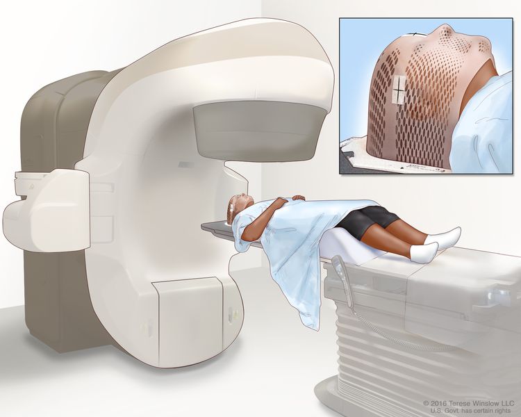
Radioterapia de haz externo dirigida al cerebro: se utiliza una máquina para administrar radiación de alta energía, que puede girar alrededor del paciente y aplicar el tratamiento desde varios ángulos. Una máscara de malla ayuda a mantener la cabeza del paciente inmóvil durante el procedimiento. Se colocan pequeñas marcas de tinta en la máscara, que se utilizan para alinear la máquina de radiación en la misma posición antes de cada sesión.
Ciertas formas de administrar radioterapia externa pueden ayudar a evitar que la radiación dañe el tejido sano cercano. Estos tipos de radioterapia son:
- Radioterapia conformacional: se utiliza una computadora para crear una imagen tridimensional (3D) del tumor y adaptar los haces de radiación a la forma del tumor.
- Radioterapia de intensidad modulada (IMRT): la IMRT es un tipo de radioterapia tridimensional (3D) en la que se utiliza una computadora para generar imágenes del tamaño y la forma del tumor. Se administran rayos finos de radiación de diferentes intensidades (fuerzas) al tumor desde muchos ángulos.
- Radiocirugía estereotáctica: la radiocirugía estereotáctica utiliza un marco rígido para la cabeza que se fija al cráneo para mantener la cabeza quieta durante el tratamiento de radiación. Una máquina dirige una única dosis grande de radiación directamente al tumor. Este procedimiento no implica cirugía. También se denomina radiocirugía estereotáxica, radiocirugía y cirugía por radiación.
Quimioterapia
La quimioterapia es un tratamiento contra el cáncer que utiliza fármacos para detener el crecimiento de las células cancerosas, ya sea destruyéndolas o impidiendo su división. Cuando la quimioterapia se administra por vía oral o se inyecta en una vena o músculo, los fármacos entran en el torrente sanguíneo y pueden llegar a las células cancerosas en todo el cuerpo ( quimioterapia sistémica ). Aunque la mayoría no pueden, algunos fármacos de quimioterapia pueden atravesar la barrera hematoencefálica y llegar a las células tumorales en el cerebro. La quimioterapia que se administra directamente en el líquido cefalorraquídeo se denomina quimioterapia intratecal . Cuando la quimioterapia se administra en un órgano, como el cerebro, o en una cavidad corporal, los fármacos afectan principalmente a las células cancerosas en esas áreas ( quimioterapia regional ).
Para tratar los tumores cerebrales, se puede usar un disco que se disuelve para administrar un medicamento de quimioterapia directamente al sitio del tumor cerebral después de que ha sido extirpado mediante cirugía. La forma en que se administra la quimioterapia depende del tipo y grado del tumor y de su ubicación en el cerebro.
See Drugs Approved for Brain Tumors for more information.
Terapia dirigida
La terapia dirigida es un tipo de tratamiento que utiliza medicamentos u otras sustancias para identificar y atacar células cancerosas específicas.
- Anticuerpo monoclonal terapia: los anticuerpos monoclonales son proteínas del sistema inmunitario proteínas made in the laboratory to treat many diseases, including cancer. As a cancer treatment, these anticuerpos se adhieren a un objetivo específico en las células cancerosas u otras células que podrían estar causando el crecimiento del cáncer. De esta forma, los anticuerpos pueden eliminar las células cancerosas, bloquear su crecimiento o evitar que se propaguen. Los anticuerpos monoclonales son administrados por infusión. Pueden utilizarse solos o para transportar medicamentos, toxinaso radioactivo material directamente a las células cancerosas.
El bevacizumab es un anticuerpo monoclonal que se une a una proteína llamada factor de crecimiento endotelial vascular (VEGF) y puede prevenir el crecimiento de nuevos vasos sanguíneos que necesitan los tumores para crecer. El bevacizumab se utiliza en el tratamiento del glioblastoma recidivante.
Se están estudiando otros tipos de terapias dirigidas para tumores cerebrales en adultos, como los inhibidores de tirosina quinasa y nuevos inhibidores de VEGF.
See Drugs Approved for Brain Tumors for more information.
Se ofrece atención de apoyo para aliviar los problemas causados por la enfermedad o su tratamiento.
Esta terapia controla los problemas o efectos secundarios causados por la enfermedad o su tratamiento y mejora la calidad de vida . En el caso de los tumores cerebrales, los cuidados de apoyo incluyen medicamentos para controlar las convulsiones y la acumulación de líquido o inflamación en el cerebro.
New types of treatment are being tested in clinical trials.
En esta sección del resumen se presentan nuevos tratamientos que se están estudiando en ensayos clínicos, pero es posible que no mencione todos ellos. La información sobre ensayos clínicos está disponible en el sitio web del NCI.
Radioterapia con haz de protones
La radioterapia con haz de protones es un tipo de radioterapia externa de alta energía que utiliza haces de protones (partículas diminutas con carga positiva) para destruir las células tumorales. Este tratamiento reduce el daño por radiación en los tejidos sanos cercanos al tumor. Se utiliza para tratar cánceres de cabeza, cuello y columna vertebral, así como de órganos como el cerebro, el ojo, el pulmón y la próstata . La radioterapia con haz de protones es diferente de la radioterapia con rayos X.
Inmunoterapia
La inmunoterapia es un tratamiento en el que se utiliza el sistema inmunológico del paciente para combatir el cáncer. Las sustancias producidas por el cuerpo o en un laboratorio se utilizan para estimular, dirigir o restaurar las defensas naturales del cuerpo contra el cáncer.
La inmunoterapia está en estudio para el tratamiento de algunos tipos de tumores cerebrales. Los tratamientos pueden ser:
- Terapia con vacunas de células dendríticas
- Terapia génica.
El tratamiento de los tumores del sistema nervioso central en adultos puede producir efectos secundarios.
For information about side effects caused by treatment for cancer, visit our Side Effects page.
Patients may want to think about taking part in a clinical trial.
For some patients, taking part in a clinical trial may be the best treatment choice. Clinical trials are part of the cancer research process. Clinical trials are done to find out if new cancer treatments are safe and effective or better than the standard treatment.
Many of today's standard treatments for cancer are based on earlier clinical trials. Patients who take part in a clinical trial may receive the standard treatment or be among the first to receive a new treatment.
Patients who take part in clinical trials also help improve the way cancer will be treated in the future. Even when clinical trials do not lead to effective new treatments, they often answer important questions and help move research forward.
Patients can enter clinical trials before, during, or after starting their cancer treatment.
Some clinical trials only include patients who have not yet received treatment. Other trials test treatments for patients whose cancer has not gotten better. There are also clinical trials that test new ways to stop cancer from recurring (coming back) or reduce the side effects of cancer treatment.
Clinical trials are taking place in many parts of the country. Information about clinical trials supported by NCI can be found on NCI’s clinical trials search webpage. Clinical trials supported by other organizations can be found on the ClinicalTrials.gov website.
Pueden ser necesarias pruebas de seguimiento.
A medida que avanza el tratamiento, se le realizarán pruebas o controles de seguimiento. Es posible que se repitan algunas pruebas para diagnosticar o estadificar el cáncer con el fin de evaluar cómo está funcionando el tratamiento. Las decisiones sobre si continuar, modificar o suspender el tratamiento pueden basarse en los resultados de estas pruebas.
Algunas pruebas seguirán realizándose de manera periódica después de terminar el tratamiento. Los resultados pueden indicar si su afección ha cambiado o si el cáncer ha redicivado (regresado).
Se pueden usar las siguientes pruebas y procedimientos para verificar si un tumor cerebral ha regresado después del tratamiento:
- Tomografía computarizada por emisión monofotónica (SPECT): es un procedimiento para detectar células tumorales malignas en el cerebro. Se inyecta una pequeña cantidad de una sustancia radiactiva en una vena o se inhala por la nariz. A medida que la sustancia circula por la sangre, una cámara gira alrededor de la cabeza y toma imágenes del cerebro. Una computadora utiliza las imágenes para crear una imagen tridimensional (3D) del cerebro. Se observará un aumento del flujo sanguíneo y mayor actividad en las zonas donde crecen las células cancerosas. Estas áreas se verán más brillantes en la imagen.
- Tomografía por emisión de positrones (PET): es un procedimiento que se utiliza para detectar células tumorales malignas en el cuerpo. Se inyecta una pequeña cantidad de glucosa (azúcar) radiactiva en una vena. El escáner gira alrededor del cuerpo y crea una imagen de dónde se usa la glucosa en el cerebro. Las células tumorales malignas aparecen más brillantes en la imagen porque son más activas y absorben más glucosa que las células normales.
Tomografía por emisión de positrones (PET, por sus siglas en inglés). El paciente se acuesta en una mesa que se desliza a través de la máquina PET. El reposacabezas y la correa blanca ayudan al paciente a permanecer quieto. Se inyecta una pequeña cantidad de glucosa radiactiva (azúcar) en la vena del paciente y un escáner genera una imagen de dónde se usa la glucosa en el cuerpo. Las células cancerosas aparecen más brillantes en la imagen porque absorben más glucosa que las células sanas.
Tratamiento del tumor cerebral primario en adultos por tipo de tumor
For information about the treatments listed below, see the Treatment Option Overview section.
Tumores astrocíticos
Gliomas de tronco cerebral
El tratamiento de los gliomas del tronco encefálico puede incluir radioterapia .
Puede utilizar la búsqueda de ensayos clínicos y encontrar ensayos clínicos sobre cáncer patrocinados por el NCI que acepten participantes. La búsqueda le permite filtrar los ensayos según el tipo de cáncer, la edad y el lugar donde se realizan los ensayos. También encontrará información general sobre los ensayos clínicos.
Tumores astrocíticos pineales
El tratamiento de los tumores astrocíticos pineales puede incluir cirugía y radioterapia . En el caso de tumores de alto grado, también se puede administrar quimioterapia .
Puede utilizar la búsqueda de ensayos clínicos y encontrar ensayos clínicos sobre cáncer patrocinados por el NCI que acepten participantes. La búsqueda le permite filtrar los ensayos según el tipo de cáncer, la edad y el lugar donde se realizan los ensayos. También encontrará información general sobre los ensayos clínicos.
Astrocitomas pilocíticos
El tratamiento de los astrocitomas pilocíticos puede incluir cirugía para extirpar el tumor. También se puede administrar radioterapia si queda algún resto del tumor después de la cirugía.
Puede utilizar la búsqueda de ensayos clínicos y encontrar ensayos clínicos sobre cáncer patrocinados por el NCI que acepten participantes. La búsqueda le permite filtrar los ensayos según el tipo de cáncer, la edad y el lugar donde se realizan los ensayos. También encontrará información general sobre los ensayos clínicos.
Astrocitomas difusos
El tratamiento de los astrocitomas difusos puede consistir en:
- Cirugía con o sin radioterapia
- Cirugía seguida de radioterapia y quimioterapia
Puede utilizar la búsqueda de ensayos clínicos y encontrar ensayos clínicos sobre cáncer patrocinados por el NCI que acepten participantes. La búsqueda le permite filtrar los ensayos según el tipo de cáncer, la edad y el lugar donde se realizan los ensayos. También encontrará información general sobre los ensayos clínicos.
Astrocitomas anaplásicos
El tratamiento de los astrocitomas anaplásicos puede consistir en:
- Cirugía y radioterapia . También se puede administrar quimioterapia .
- Cirugía y quimioterapia
- Un ensayo clínico de quimioterapia administrada en el cerebro durante una cirugía
- Un ensayo clínico de un nuevo tratamiento como complemento de un tratamiento estándar
Puede utilizar la búsqueda de ensayos clínicos y encontrar ensayos clínicos sobre cáncer patrocinados por el NCI que acepten participantes. La búsqueda le permite filtrar los ensayos según el tipo de cáncer, la edad y el lugar donde se realizan los ensayos. También encontrará información general sobre los ensayos clínicos.
Glioblastomas
El tratamiento de los glioblastomas puede consistir en:
- Cirugía seguida de radioterapia y quimioterapia administradas al mismo tiempo, seguidas de quimioterapia sola.
- Cirugía seguida de radioterapia
- Quimioterapia en el cerebro durante la cirugía
- Radioterapia y quimioterapia administradas al mismo tiempo
- Un ensayo clínico de un nuevo tratamiento como complemento de un tratamiento estándar
Puede utilizar la búsqueda de ensayos clínicos y encontrar ensayos clínicos sobre cáncer patrocinados por el NCI que acepten participantes. La búsqueda le permite filtrar los ensayos según el tipo de cáncer, la edad y el lugar donde se realizan los ensayos. También encontrará información general sobre los ensayos clínicos.
Tumores oligodendrogliales
El tratamiento de los oligodendrogliomas puede incluir cirugía con o sin radioterapia . La quimioterapia puede administrarse después de la radioterapia.
El tratamiento del oligodendroglioma anaplásico puede consistir en:
- Cirugía seguida de radioterapia con o sin quimioterapia
- Un ensayo clínico de un nuevo tratamiento como complemento de un tratamiento estándar
Puede utilizar la búsqueda de ensayos clínicos y encontrar ensayos clínicos sobre cáncer patrocinados por el NCI que acepten participantes. La búsqueda le permite filtrar los ensayos según el tipo de cáncer, la edad y el lugar donde se realizan los ensayos. También encontrará información general sobre los ensayos clínicos.
Gliomas mixtos
El tratamiento de los gliomas mixtos puede incluir cirugía y radioterapia . En ocasiones, también se administra quimioterapia .
Puede utilizar la búsqueda de ensayos clínicos y encontrar ensayos clínicos sobre cáncer patrocinados por el NCI que acepten participantes. La búsqueda le permite filtrar los ensayos según el tipo de cáncer, la edad y el lugar donde se realizan los ensayos. También encontrará información general sobre los ensayos clínicos.
Tumores ependimarios
El tratamiento de los ependimomas de grado I y II puede incluir cirugía para extirpar el tumor. También se puede administrar radioterapia si queda algún resto tumoral después de la cirugía.
El tratamiento del ependimoma anaplásico de grado 3 puede consistir en cirugía y radioterapia.
Puede utilizar la búsqueda de ensayos clínicos y encontrar ensayos clínicos sobre cáncer patrocinados por el NCI que acepten participantes. La búsqueda le permite filtrar los ensayos según el tipo de cáncer, la edad y el lugar donde se realizan los ensayos. También encontrará información general sobre los ensayos clínicos.
Meduloblastomas
El tratamiento de los meduloblastomas puede consistir en:
- Cirugía y radioterapia del cerebro y la columna vertebral .
- Un ensayo clínico que combina quimioterapia con cirugía y radioterapia en el cerebro y la columna vertebral.
Puede utilizar la búsqueda de ensayos clínicos y encontrar ensayos clínicos sobre cáncer patrocinados por el NCI que acepten participantes. La búsqueda le permite filtrar los ensayos según el tipo de cáncer, la edad y el lugar donde se realizan los ensayos. También encontrará información general sobre los ensayos clínicos.
Tumores del parénquima pineal
El tratamiento de los tumores del parénquima pineal puede incluir lo siguiente:
- Para los pineocitomas, cirugía y radioterapia .
- Para los pineoblastomas, cirugía, radioterapia y quimioterapia
Puede utilizar la búsqueda de ensayos clínicos y encontrar ensayos clínicos sobre cáncer patrocinados por el NCI que acepten participantes. La búsqueda le permite filtrar los ensayos según el tipo de cáncer, la edad y el lugar donde se realizan los ensayos. También encontrará información general sobre los ensayos clínicos.
Tumores meníngeos
El tratamiento de los meningiomas de grado 1 puede consistir en:
- Vigilancia activa de tumores sin signos ni síntomas .
- Cirugía para extirpar el tumor. Si queda algún resto del tumor después de la cirugía, también se puede administrar radioterapia .
- Radiocirugía estereotáxica para tumores que miden menos de 3 centímetros
- Radioterapia para tumores que no se pueden extirpar mediante cirugía.
El tratamiento de los meningiomas y hemangiopericitomas de grado 2 y 3 puede consistir en:
- Cirugía y radioterapia
Puede utilizar la búsqueda de ensayos clínicos y encontrar ensayos clínicos sobre cáncer patrocinados por el NCI que acepten participantes. La búsqueda le permite filtrar los ensayos según el tipo de cáncer, la edad y el lugar donde se realizan los ensayos. También encontrará información general sobre los ensayos clínicos.
Tumores de células germinales
No existe un tratamiento estándar para los tumores de células germinales (germinoma, carcinoma embrionario, coriocarcinoma y teratoma ). El tratamiento depende del aspecto de las células tumorales al microscopio, de los marcadores tumorales, de la ubicación del tumor en el cerebro y de si se puede extirpar mediante cirugía.
Puede utilizar la búsqueda de ensayos clínicos y encontrar ensayos clínicos sobre cáncer patrocinados por el NCI que acepten participantes. La búsqueda le permite filtrar los ensayos según el tipo de cáncer, la edad y el lugar donde se realizan los ensayos. También encontrará información general sobre los ensayos clínicos.
Craneofaringiomas
El tratamiento de los craneofaringiomas puede consistir en:
- Cirugía para extirpar el tumor completo
- Cirugía para extirpar la mayor cantidad posible de tumor, seguida de radioterapia
Puede utilizar la búsqueda de ensayos clínicos y encontrar ensayos clínicos sobre cáncer patrocinados por el NCI que acepten participantes. La búsqueda le permite filtrar los ensayos según el tipo de cáncer, la edad y el lugar donde se realizan los ensayos. También encontrará información general sobre los ensayos clínicos.
Tratamiento de los tumores primarios de la médula espinal en adultos
For information about the treatments listed below, see the Treatment Option Overview section.
El tratamiento de los tumores de la médula espinal puede incluir lo siguiente:
- Cirugía para extirpar el tumor
- Radioterapia.
- Participación en el ensayo clínico de un tratamiento nuevo
Tratamiento de los tumores recidivantes del sistema nervioso central en adultos
For information about the treatments listed below, see the Treatment Option Overview section.
No existe un tratamiento estándar para los tumores recurrentes del sistema nervioso central (SNC) . El tratamiento depende del estado del paciente, los posibles efectos secundarios, la ubicación del tumor en el SNC y la posibilidad de extirparlo quirúrgicamente. El tratamiento puede incluir lo siguiente:
- Quimioterapia administrada en el cerebro durante la cirugía
- Quimioterapia con medicamentos no utilizados para tratar el tumor original
- Terapia dirigida para el glioblastoma recidivante
- Radioterapia.
- Cirugía para extirpar el tumor
- Participación en el ensayo clínico de un tratamiento nuevo
Puede utilizar la búsqueda de ensayos clínicos y encontrar ensayos clínicos sobre cáncer patrocinados por el NCI que acepten participantes. La búsqueda le permite filtrar los ensayos según el tipo de cáncer, la edad y el lugar donde se realizan los ensayos. También encontrará información general sobre los ensayos clínicos.
Tratamiento de tumores cerebrales metastásicos en adultos
For information about the treatments listed below, see the Treatment Option Overview section.
El tratamiento de uno a cuatro tumores que se han diseminado al cerebro desde otra parte del cuerpo puede consistir en:
- Radioterapia en todo el cerebro con o sin cirugía.
- Radioterapia a todo el cerebro con o sin radiocirugía estereotáxica
- Radiocirugía estereotáctica
- Quimioterapia, si el tumor primario responde a los fármacos anticancerígenos . Puede combinarse con radioterapia.
El tratamiento de los tumores que se han diseminado a las leptomeninges puede consistir en:
- Quimioterapia (sistémica y/o intratecal). También se puede administrar radioterapia.
- Atención de apoyo.
Puede utilizar la búsqueda de ensayos clínicos y encontrar ensayos clínicos sobre cáncer patrocinados por el NCI que acepten participantes. La búsqueda le permite filtrar los ensayos según el tipo de cáncer, la edad y el lugar donde se realizan los ensayos. También encontrará información general sobre los ensayos clínicos.
Más información sobre los tumores del sistema nervioso central en adultos
Para obtener más información del Instituto Nacional del Cáncer sobre los tumores del sistema nervioso central (SNC) en adultos, consulte lo siguiente:
- Página de inicio del cáncer de cerebro
- NCI-CONNECT (Red integral de oncología que evalúa tumores raros del SNC)
- Medicamentos aprobados para tumores cerebrales
- Terapia dirigida para tratar el cáncer
- Immunotherapy to Treat Cancer
For general cancer information and other resources from the National Cancer Institute, visit:
Sobre este resumen del PDQ
Acerca del PDQ
El Physician Data Query (PDQ) es la base de datos integral sobre el cáncer del National Cancer Institute (NCI). La base de datos del PDQ contiene resúmenes con la última información publicada sobre prevención, detección, genética, tratamiento, atención médica de apoyo y medicina complementaria y alternativa relacionada con el cáncer. La mayoría de los resúmenes se presentan en dos versiones. Las versiones para profesionales de la salud contienen información detallada escrita en lenguaje técnico. Las versiones para pacientes están escritas en un lenguaje fácil de entender y no tan técnico. Ambas versiones contienen información precisa y actualizada sobre el cáncer. La mayoría de las versiones también están disponibles en español.
El PDQ es un servicio del NCI. El NCI es parte de los Institutos Nacionales de Salud (NIH), que son el centro de investigación biomédica del Gobierno federal. Los resúmenes del PDQ se basan en una revisión independiente de la literatura médica. No son declaraciones de políticas del NCI ni de los NIH.
Propósito de este resumen
Este resumen del PDQ sobre el cáncer contiene información actualizada sobre el tratamiento de los tumores del sistema nervioso central en adultos. Su propósito es informar y ayudar a los pacientes, las familias y los cuidadores. No da pautas ni recomendaciones formales para tomar decisiones relacionadas con la atención médica.
Revisores y actualizaciones
Los comités editoriales escriben los resúmenes de información sobre el cáncer del PDQ y los mantienen actualizados. Estos comités están formados por equipos de especialistas en el tratamiento del cáncer y otras especialidades relacionadas con esta enfermedad. Los resúmenes se revisan periódicamente y se modifican cuando hay información nueva. La fecha de actualización al pie de cada resumen indica cuándo se realizó el cambio más reciente.
The information in this patient summary was taken from the health professional version, which is reviewed regularly and updated as needed, by the PDQ Adult Treatment Editorial Board.
Información sobre ensayos clínicos
Un ensayo clínico es un estudio para responder a una pregunta científica como, por ejemplo, si un tratamiento es mejor que otro. Los ensayos se basan en estudios anteriores y en lo aprendido en el laboratorio. Cada ensayo responde a determinadas preguntas científicas que permiten encontrar nuevas y mejores formas de ayudar a los pacientes con cáncer. Durante los ensayos clínicos de tratamiento, se recopila información sobre los efectos de un nuevo tratamiento y su eficacia. Si un ensayo clínico demuestra que un nuevo tratamiento es mejor que uno que se utiliza actualmente, el nuevo tratamiento puede convertirse en “estándar”. Los pacientes pueden valorar la posibilidad de participar en un ensayo clínico. Algunos ensayos clínicos solo están abiertos a pacientes que no hayan iniciado el tratamiento.
Los ensayos clínicos se pueden encontrar en línea en el sitio web del NCI. Para obtener más información, llame al Servicio de Información sobre el Cáncer (CIS, por sus siglas en inglés), el centro de contacto del NCI, al 1-800-4-CANCER (1-800-422-6237).
Permiso de uso de este resumen
Physician Data Query (PDQ) es una marca registrada. Se autoriza el libre uso del contenido de los documentos del PDQ como texto. Sin embargo, no se podrá identificar como un resumen de información sobre cáncer del PDQ del NCI, salvo que se reproduzca en su totalidad y se actualice con regularidad. Por otra parte, se permite que los autores incluyan una oración como “en el resumen del PDQ del NCI sobre la prevención del cáncer de mama se describen, de manera concisa, los siguientes riesgos: [incluir fragmento del resumen]”.
La forma recomendada para citar este resumen del PDQ es:
Comité editorial del PDQ® sobre el tratamiento para adultos. Tratamiento de tumores del sistema nervioso central en adultos (PDQ). Bethesda, MD: National Cancer Institute. Actualizado el [DD/MM/AAAA]
Las imágenes de este resumen se utilizan con el permiso del autor, artista y/o editorial para uso exclusivo en los resúmenes del PDQ. Si desea usar una imagen de un resumen del PDQ sin incluir el resumen completo, debe obtener autorización del propietario. El National Cancer Institute no puede otorgar dicho permiso. Para obtener más información sobre el uso de las imágenes de este resumen o de otras ilustraciones relacionadas con el cáncer, consulte Visuals Online, una colección de más de 3,000 imágenes científicas.
Descargo de responsabilidad
La información de estos resúmenes no debe utilizarse para tomar decisiones sobre reembolsos de seguros. Puede encontrar más información sobre la cobertura de seguros en Cancer.gov en el sitio Manejo de la atención del cáncer.
Contáctenos
Puede encontrar más información sobre cómo contactarnos o recibir ayuda en el sitio web Cancer.gov en la página Comuníquese con el NCI. También puede enviar sus preguntas a Cancer.gov en el apartado Escríbanos del sitio web.
Actualizado:
URL de origen: https://www.cancer.gov/node/1040/syndication
Agencia de origen: National Cancer Institute (NCI)
Fecha de captura: 2013-09-14 09:00:10.0