


Non-Small Cell Lung Cancer Treatment
Obtenga una atención excepcional para el cáncer de pulmón de células no pequeñas en Montefiore Einstein Comprehensive Cancer Center. Somos uno de los primeros centros de cáncer designados por el NCI y, durante más de 50 años, hemos sido líderes en la investigación, el diagnóstico y el tratamiento de más de 200 tipos de cáncer.
Ofrecemos inmunoterapias y terapias dirigidas. Además, nuestros pacientes pueden hablar con nuestro equipo sobre la posibilidad de participar en ensayos clínicos en fase inicial, en los que pueden recibir los tratamientos más novedosos mucho antes de que estén disponibles en cualquier otro lugar.
En Montefiore Einstein Comprehensive Cancer Center, nuestra investigación está a la vanguardia de estudios innovadores y avances transformadores en tratamientos para el cáncer. Juntos trabajamos para impulsar la investigación clínica y traslacional, acelerando el ritmo en que los descubrimientos se convierten en tratamientos y terapias que realmente beneficien a los pacientes. Nuestros equipos han investigado diversos usos de las tomografías por emisión de positrones (PET, por sus siglas en inglés) realizadas antes del tratamiento. Estas exploraciones permiten identificar lesiones con alto riesgo de progresión tras la quimiorradioterapia concurrente, específicamente en casos de cáncer de pulmón de células no pequeñas localmente avanzado. También hemos explorado la inmunoterapia basada en el ligando FLT3 para pacientes cuyo cáncer de pulmón de células no pequeñas ha progresado tras el tratamiento estándar. Otro enfoque de investigación activa en nuestro centro es el desarrollo de nuevos agentes activadores de células T. Estos agentes están diseñados para dirigirse con mayor precisión a los tumores y, al mismo tiempo, causar menos efectos secundarios.
Aquí recibirá una atención compasiva y personalizada que cumple con los más altos estándares de calidad y seguridad. El tratamiento del cáncer de pulmón de células no pequeñas de Montefiore Einstein Comprehensive Cancer Center es uno de los más completos del país. Estamos comprometidos con la atención integral de la persona y ofrecemos a los pacientes una amplia variedad de servicios de apoyo y programas de cuidados paliativos para satisfacer las complejas necesidades médicas, emocionales y espirituales de los pacientes y sus familias.
Si necesita atención médica, confíe en nuestros profesionales, que se dedican con pasión a acabar con el cáncer y a atender todas sus necesidades de salud.

El Montefiore Einstein Comprehensive Cancer Center, designado como centro integral del cáncer por el National Cancer Institute (NCI), apoya la misión y las normas del NCI. La siguiente información sobre los tipos de cáncer, prevención y tratamientos ha sido facilitada por el NCI.
Tratamiento del cáncer de pulmón de células no pequeñas (PDQ®): versión para pacientes
Información general sobre el cáncer de pulmón de células no pequeñas
Puntos clave
- El cáncer de pulmón de células no pequeñas es un tipo de cáncer que se forma en los tejidos del pulmón.
- Existen varios tipos de cáncer de pulmón de células no pequeñas.
- Fumar es el principal factor de riesgo para desarrollar cáncer de pulmón de células no pequeñas.
- Entre los signos y síntomas comunes se incluyen tos y dificultad para respirar.
- Para diagnosticar y estadificar el cáncer de pulmón de células no pequeñas se utilizan pruebas que examinan los pulmones.
- Si se sospecha que tiene cáncer de pulmón, se le realizará una biopsia.
- Una vez diagnosticado el cáncer de pulmón de células no pequeñas, se realizan pruebas para determinar si las células cancerosas se han diseminado dentro del tórax o a otras partes del cuerpo.
- Some people decide to get a second opinion.
- Certain factors affect the prognosis (chance of recovery) and treatment options.
El cáncer de pulmón de células no pequeñas es un tipo de cáncer que se forma en los tejidos del pulmón.
Los pulmones son un par de órganos respiratorios con forma de cono situados en el pecho. Los pulmones introducen oxígeno en el cuerpo al inspirar. Al exhalar, liberan dióxido de carbono, un producto de desecho de las células del cuerpo. Cada pulmón tiene unas secciones llamadas lóbulos. El pulmón izquierdo tiene dos lóbulos. El pulmón derecho es ligeramente más grande y tiene tres lóbulos. Dos tubos llamados bronquios conducen desde la tráquea (tubo respiratorio) hasta los pulmones derecho e izquierdo. El cáncer de pulmón también puede formarse en los bronquios. El interior de los pulmones está formado por pequeños sacos de aire llamados alvéolos y pequeños tubos llamados bronquiolos.

Anatomy of the respiratory system showing the trachea, the right and left lungs and their lobes, and the bronchi. The lymph nodes and the diaphragm are also shown. Oxygen is inhaled into the lungs and passes through the alveoli (the tiny air sacs at the end of the bronchioles) and into the bloodstream (see inset), where it travels to the tissues throughout the body.
Una fina membrana llamada pleura recubre la parte externa de cada pulmón y la pared interna de la cavidad torácica. Esto forma un saco llamado cavidad pleural . La cavidad pleural normalmente contiene una pequeña cantidad de líquido que ayuda a que los pulmones se muevan con fluidez dentro del tórax al respirar.
Existen dos tipos principales de cáncer de pulmón: el cáncer de pulmón de células no pequeñas y el cáncer de pulmón de células pequeñas. El cáncer de pulmón de células no pequeñas es más común que el cáncer de pulmón de células pequeñas.
Existen varios tipos de cáncer de pulmón de células no pequeñas.
Cada tipo de cáncer de pulmón de células no pequeñas tiene diferentes tipos de células cancerosas. Las células cancerosas de cada tipo crecen y se diseminan de diferentes maneras. Los tipos de cáncer de pulmón de células no pequeñas se denominan según los tipos de células que se encuentran en el cáncer y cómo se ven las células con microscopio:
- El carcinoma escamocelular es un tipo de cáncer de pulmón que se forma en las células delgadas y planas que recubren el interior de los pulmones. También se denomina carcinoma epidermoide.
- El carcinoma de células grandes es un tipo de cáncer de pulmón que puede comenzar en varios tipos de células grandes.
- El adenocarcinoma es un tipo de cáncer de pulmón que comienza en las células que recubren los alvéolos y producen sustancias como la mucosidad.
Los tipos menos comunes de cáncer de pulmón de células no pequeñas incluyen el carcinoma adenoescamoso, el carcinoma sarcomatoide, el carcinoma de las glándulas salivales, el tumor carcinoide y el carcinoma no clasificado.
Fumar es el principal factor de riesgo para desarrollar cáncer de pulmón de células no pequeñas.
El cáncer de pulmón se debe a ciertos cambios en el funcionamiento de las células pulmonares, particularmente en su crecimiento y división anómala. Existen muchos factores de riesgo para el cáncer de pulmón, pero no todos lo causan directamente. En cambio, estos factores aumentan la probabilidad de daño al ADN celular, lo que puede derivar en cáncer de pulmón. Puede encontrar más información sobre cómo se desarrolla esta enfermedad en la sección ¿Qué es el cáncer?
Un factor de riesgo es cualquier elemento que incrementa la probabilidad de desarrollar una enfermedad. Algunos factores de riesgo del cáncer de pulmón, como el tabaquismo, se pueden modificar. Sin embargo, los factores de riesgo también abarcan aspectos que no se pueden cambiar, como la genética, la edad y los el historial familiar. Conocer los factores de riesgo del cáncer de pulmón puede ayudarle a realizar cambios que podrían disminuir el riesgo de desarrollarlo.
Fumar tabaco ahora o en el pasado es el factor de riesgo más importante para el cáncer de pulmón. Fumar cigarrillos, pipas o puros aumenta el riesgo de cáncer de pulmón. Cuanto antes se empieza a fumar, cuanto más se fuma y cuantos más años se fuma, mayor es el riesgo de cáncer de pulmón.
Otros factores de riesgo para el cáncer de pulmón incluyen:
- Estar expuesto al humo de tabaco
- Estar expuesto al asbesto, arsénico, cromo, berilio, níquel, hollín o alquitrán en el lugar de trabajo
- Estar expuesto a radiación de:
- Radioterapia en la mama o el tórax
- Radón en el hogar o en el lugar de trabajo
- Diagnóstico por imágenes como tomografías computarizadas
- Radiación de una bomba atómica
- Vivir en un lugar con contaminación atmosférica
- Tener un historial familiar de cáncer de pulmón
- Estar infectado con VIH
- Tomar suplementos de betacaroteno y ser un gran fumador
La edad avanzada es el principal factor de riesgo para la mayoría de los cánceres. La probabilidad de padecer cáncer aumenta con la edad.
Tener uno o más de estos factores de riesgo no significa necesariamente que vaya a tener cáncer de pulmón. Muchas personas con factores de riesgo nunca desarrollan cáncer de pulmón, mientras que otras sin factores de riesgo conocidos sí lo desarrollan. Hable con su médico si cree que puede tener un riesgo elevado.
Cuando el tabaquismo se combina con otros factores de riesgo, el riesgo de desarrollar cáncer de pulmón aumenta significativamente.
Entre los signos y síntomas comunes se incluyen tos y dificultad para respirar.
A veces, el cáncer de pulmón no causa ningún signo ni síntoma. Puede detectarse durante una radiografía de tórax realizada por otra afección. Los signos y síntomas pueden ser causados por el cáncer de pulmón o por otras afecciones. Consulte con su médico si presenta:
- molestias o dolor en el pecho
- Tos que no desaparece o que empeora con el tiempo
- dificultad para respirar
- sibilancias
- sangre en el esputo (moco expectorado de los pulmones)
- ronquera
- Pérdida de apetito
- weight loss for no known reason
- fatigue
- dificultad para tragar
- Hinchazón en la cara y/o venas del cuello
Para diagnosticar y estadificar el cáncer de pulmón de células no pequeñas se utilizan pruebas que examinan los pulmones.
El cáncer de pulmón de células no pequeñas suele diagnosticarse mediante pruebas y procedimientos que permiten obtener imágenes del pulmón y la zona que lo rodea. El proceso utilizado para determinar si las células cancerosas se han diseminado dentro y alrededor del pulmón se denomina estadificación. Las pruebas y los procedimientos para detectar, diagnosticar y estadificar el cáncer de pulmón de células no pequeñas suelen realizarse al mismo tiempo. Para planificar el tratamiento, es importante conocer el estadio de la enfermedad y si el cáncer puede extirparse mediante cirugía.
In addition to asking about your personal and family health history and doing a physical exam, your doctor may perform the following tests and procedures:
- Las pruebas de laboratorio son procedimientos médicos que analizan muestras de tejido, sangre, orina u otras sustancias del cuerpo. Estas pruebas ayudan a diagnosticar enfermedades, planificar y controlar tratamientos, o monitorear la enfermedad a lo largo del tiempo.
- Radiografía de tórax Es un tipo de radiación que puede atravesar el cuerpo y crear imágenes de los órganos y huesos dentro del tórax.
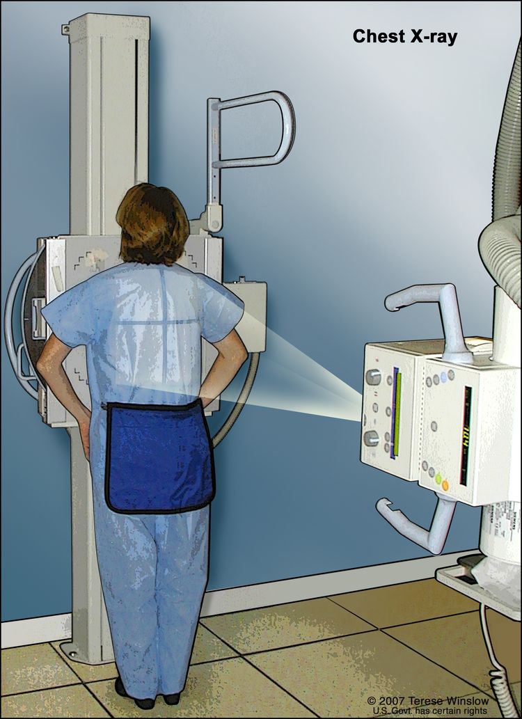
Una radiografía de tórax se utiliza para obtener imágenes de las estructuras y órganos dentro del tórax. Los rayos X atraviesan el cuerpo del paciente y se proyectan sobre una película o una computadora.
- La tomografía computarizada (TC) del cerebro, el tórax y el abdomen utiliza una computadora conectada a una máquina de rayos X para crear una serie de imágenes detalladas de áreas internas del cuerpo. Las imágenes se toman desde diferentes ángulos y se utilizan para crear vistas tridimensionales de los tejidos y órganos. Se puede inyectar un tinte en una vena o ingerirlo para ayudar a que los órganos o tejidos se vean con más claridad. Este procedimiento también se denomina tomografía axial computarizada o exploración por TAC.
Si se sospecha que tiene cáncer de pulmón, se le realizará una biopsia.
Es posible que le realicen uno de los siguientes tipos de biopsias:
- Biopsia de pulmón por aspiración con aguja fina (AAF) consiste en extraer tejido o líquido del pulmón utilizando una aguja delgada. Una tomografía computarizada, ecografía, u otro procedimiento de imagen Se utiliza para localizar tejido o líquido anormal en el pulmón. Un pequeño incisión se puede hacer en la piel donde se inserta la aguja de la biopsia en el tejido o líquido anómalo. Se extrae una muestra con la aguja y se envía al laboratorio. A continuación, un patólogo examina la muestra con un microscopio para buscar células cancerosas. Después del procedimiento, se realiza una radiografía de tórax para asegurarse de que no haya fugas de aire del pulmón al tórax.
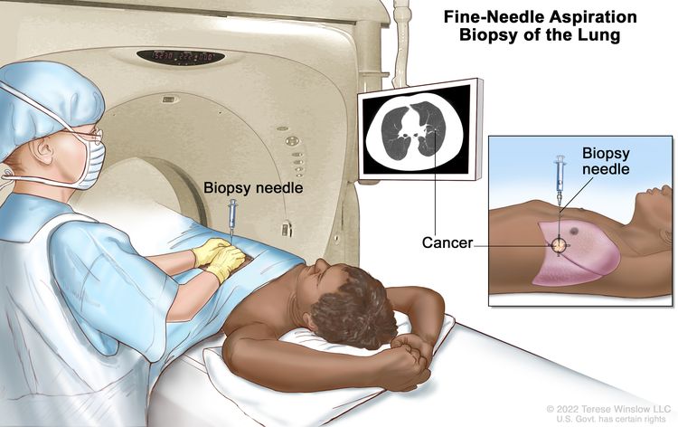
Biopsia por aspiración con aguja fina del pulmón. El paciente se recuesta sobre una mesa que se desliza a través de la máquina de tomografía computarizada (TC), la cual toma imágenes de rayos X del interior del cuerpo. Las imágenes de rayos X ayudan al médico a ver dónde se encuentra el tejido anómalo en el pulmón. Se inserta una aguja de biopsia a través de la pared torácica y en el área del tejido pulmonar anómalo. Se extrae un pequeño fragmento de tejido a través de la aguja y se examina con un microscopio para detectar signos de cáncer.
La ecografía endoscópica (EUS) es un tipo de ultrasonido que se puede utilizar para guiar una biopsia por aspiración con aguja fina (BAAF) de pulmón, ganglios linfáticos u otras áreas. La EUS es un procedimiento en el que se introduce un endoscopio en el cuerpo. Un endoscopio es un instrumento delgado, con forma de tubo, que cuenta con una luz y una lente para la visualización. Una sonda en el extremo del endoscopio se utiliza para hacer rebotar ondas sonoras de alta energía (ultrasonido) en los tejidos u órganos internos y producir ecos. Estos ecos forman una imagen de los tejidos corporales llamada ecografía .
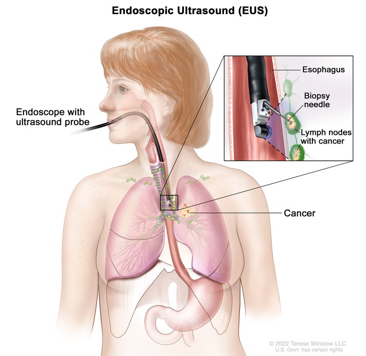
Biopsia aspirativa con aguja fina guiada por ultrasonido endoscópico. Se inserta un endoscopio que tiene una sonda de ultrasonido y una aguja de biopsia a través de la boca hasta el esófago. La sonda hace rebotar ondas de sonido en los tejidos del cuerpo para producir ecos que forman una ecografía (imagen de computadora) de los ganglios linfáticos cerca del esófago. La ecografía ayuda al médico a ver dónde colocar la aguja de biopsia para extraer tejido de los ganglios linfáticos. Este tejido se examina bajo un microscopio para detectar signos de cáncer.
- Broncoscopia Es un procedimiento para examinar el interior de la tráquea y las vías respiratorias principales de los pulmones en busca de anomalías. Se introduce un broncoscopio por la nariz o la boca hasta la tráquea y los pulmones. El broncoscopio es un instrumento delgado, con forma de tubo, que cuenta con una luz y una lente para la visualización. También puede incluir un instrumento para extraer muestras de tejido, que se examinan al microscopio en busca de signos de cáncer.
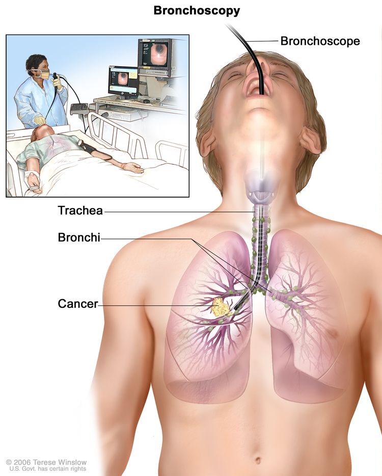
Broncoscopia. Se introduce un broncoscopio a través de la boca, la tráquea y los bronquios principales hasta el pulmón, con el objetivo de identificar áreas anómalas. El broncoscopio es un instrumento delgado en forma de tubo, con una luz y una lente para observar. En algunos casos, también cuenta con una herramienta de corte para la toma de muestras de tejido, que se analizan con microscopio en busca de posibles signos de enfermedad.
- La toracoscopia es un procedimiento quirúrgico que se realiza para examinar los órganos del interior del tórax y detectar áreas anómalas. Se realiza una incisión (corte) entre dos costillas y se inserta un toracoscopio en el tórax. Un toracoscopio es un instrumento delgado en forma de tubo con una luz y una lente para observar. También puede tener una herramienta para extraer muestras de tejido o ganglios linfáticos, que se examinan bajo un microscopio para detectar signos de cáncer. En algunos casos, este procedimiento se utiliza para extirpar parte del esófago o el pulmón. Si no se puede llegar a ciertos tejidos, órganos o ganglios linfáticos, se puede realizar una toracotomía. En este procedimiento, se realiza una incisión más grande entre las costillas y se abre el tórax.
- La toracocentesis es la extracción de líquido del espacio entre el revestimiento del tórax y el pulmón mediante una aguja. Un patólogo observa el líquido bajo un microscopio para buscar células cancerosas.
- La mediastinoscopia es un procedimiento quirúrgico para examinar los órganos, tejidos y ganglios linfáticos entre los pulmones en busca de áreas anómalas. Se realiza una incisión (corte) en la parte superior del esternón y se inserta un mediastinoscopio en el pecho. Un mediastinoscopio es un instrumento delgado, similar a un tubo, con una luz y una lente para observar. También puede tener una herramienta para extraer muestras de tejido o ganglios linfáticos, que se examinan bajo un microscopio para detectar signos de cáncer.
- La mediastinotomía anterior es un procedimiento quirúrgico para examinar los órganos y tejidos entre los pulmones y entre el esternón y el corazón en busca de áreas anómalas. Se realiza una incisión (corte) junto al esternón y se inserta un mediastinoscopio en el pecho. Un mediastinoscopio es un instrumento delgado, similar a un tubo, con una luz y una lente para observar. También puede tener una herramienta para extraer muestras de tejido o ganglios linfáticos, que se examinan bajo un microscopio para detectar signos de cáncer. Esto también se llama procedimiento de Chamberlain.
- La biopsia de ganglio linfático es la extirpación total o parcial de un ganglio linfático. Un patólogo observa el tejido del ganglio linfático bajo un microscopio para detectar la presencia de células cancerosas. La biopsia de ganglio linfático se puede realizar al mismo tiempo que otros tipos de biopsias.
Para estudiar el tejido de la biopsia se pueden realizar una o más de las siguientes pruebas de laboratorio:
- Las pruebas moleculares buscan determinados genes, proteínas u otras moléculas en una muestra de tejido, sangre u otro líquido corporal. Las pruebas moleculares buscan determinados cambios genéticos o cromosómicos que se producen en el cáncer de pulmón de células no pequeñas.
- La inmunohistoquímica utiliza anticuerpos para detectar determinados antígenos (marcadores) en una muestra de tejido de un paciente. Los anticuerpos suelen estar unidos a una enzima o a un colorante fluorescente. Una vez que los anticuerpos se unen a un antígeno específico en la muestra de tejido, la enzima o el colorante se activan y el antígeno se puede observar con un microscopio. Este tipo de prueba se utiliza para ayudar a diagnosticar el cáncer y para ayudar a diferenciar un tipo de cáncer de otro.
Una vez diagnosticado el cáncer de pulmón de células no pequeñas, se realizan pruebas para determinar si las células cancerosas se han diseminado dentro del tórax o a otras partes del cuerpo.
El proceso que se utiliza para determinar si el cáncer se ha extendido dentro del pecho o a otras partes del cuerpo se denomina estadificación. La información obtenida a partir del proceso de estadificación determina el estadio de la enfermedad. Es importante conocer el estadio para planificar el tratamiento. Algunas de las pruebas que se utilizan para diagnosticar el cáncer de pulmón de células no pequeñas también se utilizan para estadificar la enfermedad.
Las pruebas de imagen que se pueden utilizar en el proceso de estadificación incluyen:
- La resonancia magnética ( RM ) del cerebro utiliza un imán, ondas de radio y una computadora para crear una serie de imágenes detalladas de las áreas internas del cerebro. Este procedimiento también se conoce como resonancia magnética nuclear (RMN).
- La PET (tomografía por emisión de positrones) utiliza una pequeña cantidad de glucosa radiactiva (azúcar) que se inyecta en una vena. A continuación, un escáner gira alrededor del cuerpo para generar imágenes computarizadas detalladas de las áreas internas donde se absorbe la glucosa. Dado que las células cancerosas suelen absorber más glucosa que las células normales, las imágenes pueden utilizarse para detectar células cancerosas en el cuerpo. La PET y el TAC pueden realizarse simultáneamente. Esto se denomina PET-TC.
- La gammagrafía ósea detecta células cancerosas en los huesos. Se inyecta una cantidad muy pequeña de material radiactivo en una vena y este viaja a través del torrente sanguíneo. El material radiactivo se acumula en los huesos con cáncer y es detectado por un escáner.
- La prueba de función pulmonar (PFP) verifica el funcionamiento de los pulmones. Mide la cantidad de aire que pueden contener los pulmones y la rapidez con la que el aire entra y sale de ellos. También mide la cantidad de oxígeno que se utiliza y la cantidad de dióxido de carbono que se libera durante la respiración. Esto también se denomina prueba de función pulmonar.
- La aspiración y biopsia de médula ósea consiste en extraer médula ósea, sangre y un pequeño trozo de hueso mediante la inserción de una aguja hueca en el hueso de la cadera o el esternón. Un patólogo observa la médula ósea, la sangre y el hueso bajo un microscopio para buscar signos de cáncer.
Some people decide to get a second opinion.
Es posible que desee obtener una segunda opinión para confirmar su diagnóstico de cáncer de pulmón de células no pequeñas y su plan de tratamiento. Si busca una segunda opinión, deberá obtener los resultados de las pruebas médicas y los informes del primer médico para compartirlos con el segundo médico. Este revisará el informe patológico, las diapositivas y las exploraciones. Puede que esté de acuerdo con el primer médico, sugiera cambios u otro enfoque de tratamiento, o le dé más información sobre su cáncer.
Para obtener más información sobre cómo elegir un médico y obtener una segunda opinión, visite la sección «Cómo encontrar atención oncológica» . Puede comunicarse con el Servicio de Información Oncológica del NCI por chat, correo electrónico o teléfono (en inglés y español) para obtener ayuda para encontrar un médico, un hospital o una segunda opinión. Para preguntas que pueda hacer durante sus citas, visite la sección «Preguntas para hacerle a su médico sobre el cáncer» .
Certain factors affect the prognosis (chance of recovery) and treatment options.
El pronóstico y las opciones de tratamiento dependen de:
- El estadio del cáncer (el tamaño del tumor y si está sólo en el pulmón o se ha propagado a otras partes del cuerpo)
- El tipo de cáncer de pulmón
- Si el cáncer tiene mutaciones (cambios) en ciertos genes, como el gen del receptor del factor de crecimiento epidérmico (EGFR) o el gen de la quinasa del linfoma anaplásico (ALK)
- Si hay signos y síntomas como tos o dificultad para respirar
- Su salud general
Para la mayoría de las personas con cáncer de pulmón de células no pequeñas, los tratamientos actuales no curan el cáncer. Si se le diagnostica cáncer de pulmón, tal vez desee valorar la posibilidad de participar en uno de los numerosos ensayos clínicos que se están llevando a cabo para mejorar el tratamiento o la calidad de vida. Se están realizando ensayos clínicos en la mayor parte del país para personas con todos los estadios de cáncer de pulmón de células no pequeñas. Puede obtener información sobre los ensayos clínicos actuales en Información sobre ensayos clínicos para pacientes y cuidadores.
Estadios del cáncer de pulmón de células no pequeñas
Puntos clave
- Para el cáncer de pulmón de células no pequeñas se utilizan los siguientes estadios:
- Cáncer de pulmón de células no pequeñas en estadio oculto
- Estadio 0 (carcinoma in situ)
- Cáncer de pulmón de células no pequeñas en estadio I (también llamado estadio 1)
- Cáncer de pulmón de células no pequeñas en estadio II (también llamado estadio 2)
- Cáncer de pulmón de células no pequeñas en estadio III (también llamado estadio 3)
- Cáncer de pulmón de células no pequeñas en estadio IV (también llamado estadio 4)
- El cáncer de pulmón de células no pequeñas puede recidivar (regresar) después de haber sido tratado.
El estadio del cáncer describe la extensión del cáncer en el cuerpo, el tamaño del tumor, si se ha propagado y hasta dónde se ha propagado desde el lugar donde se formó originalmente. Es importante conocer el estadio del cáncer de pulmón de células no pequeñas para planificar el mejor tratamiento.
Existen varios sistemas de estadificación del cáncer que describen la extensión del cáncer. La estadificación del cáncer de pulmón de células no pequeñas suele utilizar el sistema de estadificación TNM. El cáncer puede describirse mediante este sistema de estadificación en su informe patológico. Según los resultados del TNM, se asigna un estadio (I, II, III o IV, también escrito como 1, 2, 3 o 4) a su cáncer. Cuando le informe sobre su diagnóstico, es posible que su médico describa el cáncer como uno de estos estadios.
Más sobre las pruebas para estadificar el cáncer de pulmón de células no pequeñas. Más información sobre la estadificación del cáncer.
Para el cáncer de pulmón de células no pequeñas se utilizan los siguientes estadios:
Cáncer de pulmón de células no pequeñas en estadio oculto
En el estadio oculto (escondido), el cáncer no se puede ver mediante imágenes o broncoscopia. Las células cancerosas se encuentran en el esputo o en los lavados bronquiales (una muestra de células tomadas del interior de las vías respiratorias que conducen a los pulmones). El cáncer puede haberse extendido a otras partes del cuerpo.
Estadio 0 (carcinoma in situ)
En el estadio 0, se encuentran células anómalas en el revestimiento de las vías respiratorias. Estas células anómalas pueden convertirse en cáncer y extenderse al tejido normal cercano. El estadio 0 puede ser adenocarcinoma in situ (AIS) o carcinoma de células escamosas in situ (SCIS).
Cáncer de pulmón de células no pequeñas en estadio I (también llamado estadio 1)
En la etapa I, el cáncer ya se ha formado. La etapa I se divide en etapas IA y IB.
- Estadio IA:

Cáncer de pulmón en estadio IA. El tumor está solo en el pulmón y mide 3 centímetros o menos. El cáncer no se ha propagado a los ganglios linfáticos.
El tumor se encuentra únicamente en el pulmón y mide 3 centímetros o menos. El cáncer no se ha extendido a los ganglios linfáticos .
- Estadio IB:
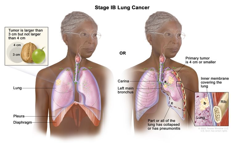
Cáncer de pulmón en estadio IB. El tumor mide más de 3 centímetros, pero no más de 4 centímetros. El cáncer no se ha extendido a los ganglios linfáticos; o el tumor mide 4 centímetros o menos. El cáncer no se ha extendido a los ganglios linfáticos y se puede dar una o más de las siguientes condiciones: (a) el cáncer se ha extendido al bronquio principal, pero no se ha extendido a la carina; y/o (b) el cáncer se ha extendido a la membrana interna que recubre el pulmón; y/o (c) parte del pulmón o todo el pulmón se ha colapsado o tiene neumonitis (inflamación del pulmón).
El tumor mide más de 3 centímetros, pero no más de 4 centímetros. El cáncer no se ha extendido a los ganglios linfáticos.
o
El tumor mide 4 centímetros o menos y se cumple al menos una de las siguientes condiciones:
- El cáncer se ha extendido al bronquio principal, pero no a la carina.
- El cáncer se ha extendido a la capa más interna de la membrana que cubre el pulmón.
- Parte del pulmón o todo el pulmón se ha colapsado o ha desarrollado neumonitis.
El cáncer no se ha extendido a los ganglios linfáticos.
Cáncer de pulmón de células no pequeñas en estadio II (también llamado estadio 2)
El estadio II se divide en los estadios IIA y IIB.
- Estadio IIA:
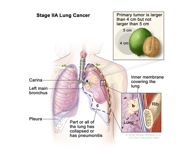
Cáncer de pulmón en estadio IIA. El tumor mide más de 4 centímetros, pero no más de 5 centímetros. El cáncer no se ha extendido a los ganglios linfáticos y se pueden dar una o más de las siguientes condiciones: (a) el cáncer se ha extendido al bronquio principal, pero no se ha extendido a la carina; y/o (b) el cáncer se ha extendido a la membrana interna que recubre el pulmón; y/o (c) parte del pulmón o todo el pulmón se ha colapsado o tiene neumonitis (inflamación del pulmón).
El tumor mide más de 4 centímetros pero no más de 5 centímetros. El cáncer no se ha diseminado a los ganglios linfáticos y se puede encontrar uno o más de los siguientes hallazgos:
- El cáncer se ha extendido al bronquio principal, pero no a la carina.
- El cáncer se ha extendido a la capa más interna de la membrana que cubre el pulmón.
- Parte del pulmón o todo el pulmón se ha colapsado o ha desarrollado neumonitis.
- Estadio IIB:
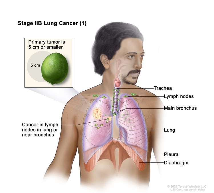
Cáncer de pulmón en estadio IIB (1). El tumor primario mide 5 centímetros o menos y el cáncer se diseminó a los ganglios linfáticos del mismo lado del tórax que el tumor primario. Los ganglios linfáticos con cáncer están en el pulmón o cerca del bronquio.
El tumor mide 5 centímetros o menos y el cáncer se ha diseminado a los ganglios linfáticos del mismo lado del tórax que el tumor primario. Los ganglios linfáticos con cáncer se encuentran en el pulmón o cerca del bronquio. Además, se puede dar una o más de las siguientes condiciones:
- El cáncer se ha extendido al bronquio principal, pero no a la carina.
- El cáncer se ha extendido a la capa más interna de la membrana que cubre el pulmón.
- Parte del pulmón o todo el pulmón se ha colapsado o ha desarrollado neumonitis.
o
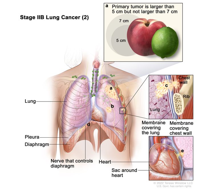
Cáncer de pulmón en estadio IIB (2). El cáncer no se ha extendido a los ganglios linfáticos y se da una o más de las siguientes condiciones: (a) el tumor primario mide más de 5 centímetros, pero no más de 7 centímetros; y/o (b) hay uno o más tumores separados en el mismo lóbulo del pulmón que el tumor primario; y/o el cáncer se ha extendido a cualquiera de los siguientes lugares: (c) la pared torácica y/o la membrana que recubre el interior de la pared torácica, (d) el nervio que controla el diafragma, y/o (e) la capa externa del tejido del saco que rodea el corazón.
El cáncer no se ha extendido a los ganglios linfáticos y se da una o más de las siguientes condiciones:
- El tumor mide más de 5 centímetros, pero no más de 7 centímetros.
- Hay uno o más tumores separados en el mismo lóbulo del pulmón que el tumor primario.
- El cáncer se ha extendido a cualquiera de los siguientes lugares:
- La membrana que recubre el interior de la pared torácica
- La pared torácica
- El nervio que controla el diafragma
- La capa exterior de tejido del saco que rodea el corazón
Cáncer de pulmón de células no pequeñas en estadio III (también llamado estadio 3)
El estadio III se divide en los estadios IIIA, IIIB y IIIC.
- Estadio IIIA:
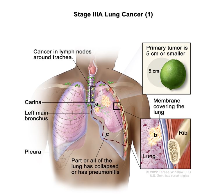
Cáncer de pulmón en estadio IIIA (1). El tumor mide 5 centímetros o menos y el cáncer se ha extendido a los ganglios linfáticos del mismo lado del tórax que el tumor primario. Los ganglios linfáticos con cáncer se encuentran alrededor de la tráquea o la aorta (no se muestran), o donde la tráquea se divide en los bronquios. Además, se puede dar una o más de las siguientes condiciones: (a) el cáncer se ha extendido al bronquio principal, pero no a la carina; y/o (b) el cáncer se ha extendido a la membrana interna que recubre el pulmón; y/o (c) parte del pulmón o todo el pulmón se ha colapsado o tiene neumonitis (inflamación del pulmón).
El tumor mide 5 centímetros o menos y el cáncer se ha diseminado a los ganglios linfáticos del mismo lado del tórax que el tumor primario . Los ganglios linfáticos afectados se encuentran alrededor de la tráquea o la aorta, o en la bifurcación de la tráquea con los bronquios . Además, se puede encontrar uno o más de los siguientes hallazgos:
- El cáncer se ha extendido al bronquio principal, pero no a la carina.
- El cáncer se ha extendido a la capa más interna de la membrana que cubre el pulmón.
- Parte del pulmón o todo el pulmón se ha colapsado o ha desarrollado neumonitis.
o
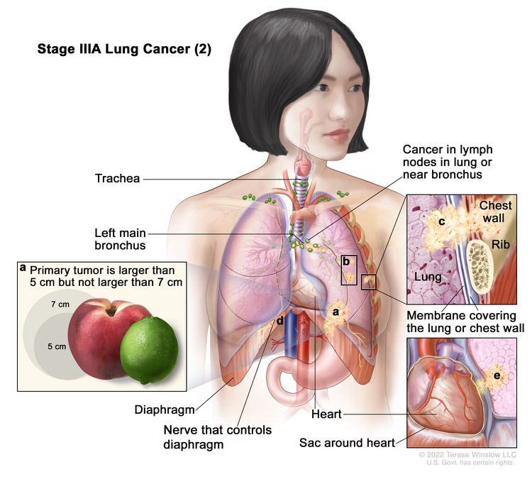
Cáncer de pulmón en estadio IIIA (2). El cáncer se ha diseminado a los ganglios linfáticos del mismo lado del tórax que el tumor primario. Los ganglios linfáticos afectados están en el pulmón o cerca del bronquio. Además, se presenta da una o más de las siguientes condiciones: (a) el tumor mide más de 5 centímetros, pero no más de 7 centímetros; y/o (b) hay uno o más tumores separados en el mismo lóbulo del pulmón que el tumor primario; y/o el cáncer se diseminó a cualquiera de los siguientes: (c) la pared torácica y/o la membrana que recubre el interior de la pared torácica, (d) el nervio que controla el diafragma y/o (e) la parte externa capa de tejido del saco que rodea el corazón.
El cáncer se ha diseminado a los ganglios linfáticos del mismo lado del tórax que el tumor primario. Los ganglios linfáticos afectados están en el pulmón o cerca del bronquio. Además, se da una o más de las siguientes condiciones:
- El tumor mide más de 5 centímetros, pero no más de 7 centímetros.
- Hay uno o más tumores separados en el mismo lóbulo del pulmón que el tumor primario.
- El cáncer se ha extendido a cualquiera de los siguientes lugares:
- La membrana que recubre el interior de la pared torácica
- La pared torácica
- El nervio que controla el diafragma
- La capa exterior de tejido del saco que rodea el corazón
o
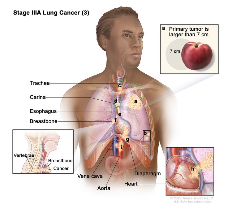
Cáncer de pulmón en estadio IIIA (3). Es posible que el cáncer se haya propagado a los ganglios linfáticos del mismo lado del tórax que el tumor primario. Los ganglios linfáticos afectados están en el pulmón o cerca del bronquio. Además, se presenta uno o más de los siguientes: (a) el tumor primario mide más de 7 centímetros; y/o (b) hay uno o más tumores separados en un lóbulo diferente del pulmón con el tumor primario; y/o el tumor es de cualquier tamaño y el cáncer se diseminó a cualquiera de los siguientes: (c) tráquea, (d) carina, (e) esófago, (f) esternón o columna vertebral, (g) diafragma, (h) corazón, (i) los principales vasos sanguíneos que conducen hacia o desde el corazón (aorta o vena cava), o el nervio que controla la laringe (no se muestra).
Es posible que el cáncer se haya propagado a los ganglios linfáticos del mismo lado del tórax que el tumor primario. Los ganglios linfáticos afectados están en el pulmón o cerca del bronquio. Además, se pueden dar una o más de las siguientes condiciones:
- El tumor mide más de 7 centímetros.
- Hay uno o más tumores separados en un lóbulo distinto del pulmón donde se encuentra el tumor primario.
- El tumor es de cualquier tamaño y el cáncer se ha extendido a cualquiera de los siguientes lugares:
- Tráquea
- La carina
- El esófago
- El esternón o columna vertebral
- El diafragma
- El corazón
- Los principales vasos sanguíneos que llegan al corazón o salen del mismo (aorta o vena cava)
- El nervio que controla la laringe (caja de la voz)
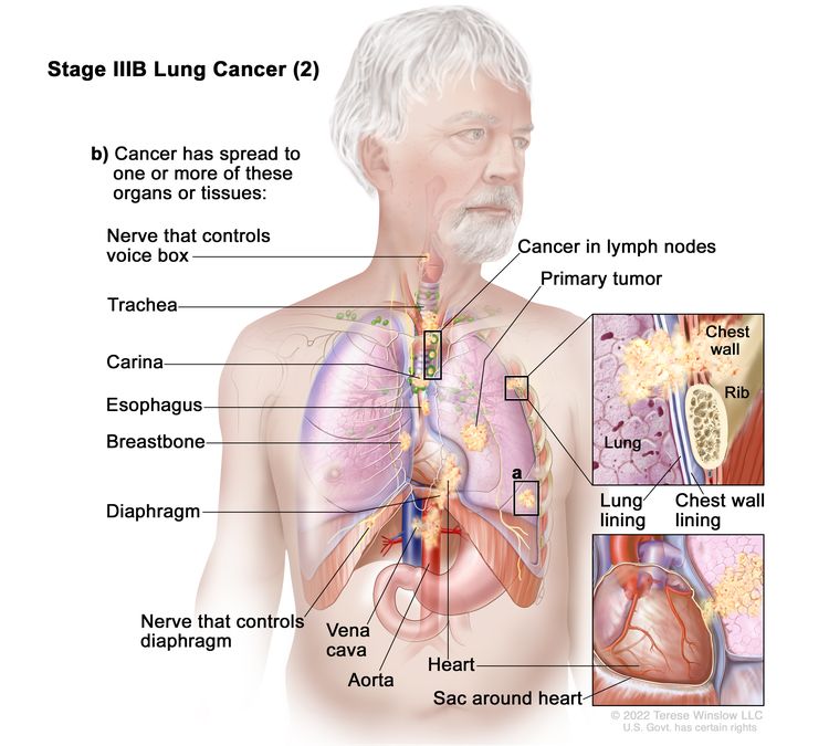
- Estadio IIIB:
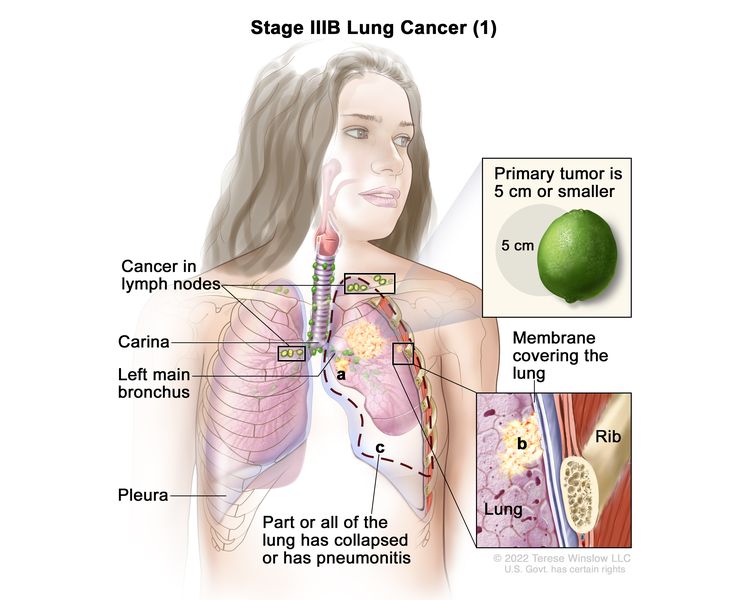
Cáncer de pulmón en estadio IIIB (1). El tumor primario mide 5 centímetros o menos y el cáncer se ha extendido a los ganglios linfáticos por encima de la clavícula en el mismo lado del tórax que el tumor primario o a cualquier ganglio linfático en el lado opuesto del tórax que el tumor primario. Además, se pueden dar uno o más de las siguientes condiciones: (a) el cáncer se ha extendido al bronquio principal, pero no se ha extendido a la carina; y/o (b) el cáncer se ha extendido a la membrana interna que cubre el pulmón; y/o (c) parte del pulmón o todo el pulmón se ha colapsado o tiene neumonitis (inflamación del pulmón).
El tumor mide 5 centímetros o menos y el cáncer se ha extendido a los ganglios linfáticos por encima de la clavícula del mismo lado del tórax que el tumor primario o a cualquier ganglio linfático del lado opuesto del tórax que el tumor primario. Además, se pueden dar una o más de las siguientes condiciones:
- El cáncer se ha extendido al bronquio principal, pero no a la carina.
- El cáncer se ha extendido a la capa más interna de la membrana que cubre el pulmón.
- Parte del pulmón o todo el pulmón se ha colapsado o ha desarrollado neumonitis.
o
Cáncer de pulmón en estadio IIIB (2). El tumor puede tener cualquier tamaño y el cáncer se diseminó a los ganglios linfáticos del mismo lado del tórax que el tumor primario. Los ganglios linfáticos con cáncer se encuentran alrededor de la tráquea o la aorta (no se muestra), o donde la tráquea se divide en los bronquios. Además, se encuentra uno o más de los siguientes: (a) hay uno o más tumores separados en el mismo lóbulo o en un lóbulo diferente del pulmón con el tumor primario; y/o (b) el cáncer se diseminó a cualquiera de los siguientes: la pared torácica o la membrana que recubre el interior de la pared torácica, el nervio que controla la laringe, la tráquea, la carina, el esófago, el esternón o columna vertebral (no se muestra), el diafragma, el nervio que controla el diafragma, el corazón, los principales vasos sanguíneos que conducen hacia o desde el corazón (aorta o vena cava), o la capa externa de tejido del saco que rodea el corazón.
El tumor puede tener cualquier tamaño y el cáncer se diseminó a los ganglios linfáticos del mismo lado del tórax que el tumor primario. Los ganglios linfáticos con cáncer se encuentran alrededor de la tráquea o la aorta, o donde la tráquea se divide en los bronquios. Además, se encuentra uno o más de los siguientes:
- Hay uno o más tumores separados en el mismo lóbulo o en un lóbulo diferente del pulmón con el tumor primario.
- El cáncer se ha extendido a cualquiera de los siguientes lugares:
- La membrana que recubre el interior de la pared torácica
- La pared torácica
- El nervio que controla el diafragma
- La capa exterior de tejido del saco que rodea el corazón
- Tráquea
- La carina
- el esófago
- El esternón o columna vertebral
- El diafragma
- El corazón
- Los principales vasos sanguíneos que llegan al corazón o salen del mismo (aorta o vena cava)
- El nervio que controla la laringe (caja de la voz)
- Estadio IIIC:

Cáncer de pulmón en estadio IIIC. El tumor puede tener cualquier tamaño y el cáncer se ha extendido a los ganglios linfáticos por encima de la clavícula en el mismo lado del tórax que el tumor primario o a cualquier ganglio linfático en el lado opuesto del tórax que el tumor primario. Además, se da una o más de las siguientes condiciones: (a) hay uno o más tumores separados en el mismo lóbulo o en un lóbulo diferente del pulmón con el tumor primario; y/o (b) el cáncer se ha extendido a cualquiera de los siguientes lugares: la pared torácica o la membrana que recubre el interior de la pared torácica, el nervio que controla la laringe, la tráquea, la carina, el esófago, el esternón o columna vertebral (no se muestra), el diafragma, el nervio que controla el diafragma, el corazón, los principales vasos sanguíneos que conducen hacia o desde el corazón (aorta o vena cava), o la capa exterior de tejido del saco que rodea el corazón.
El tumor puede tener cualquier tamaño y el cáncer se ha extendido a los ganglios linfáticos por encima de la clavícula en el mismo lado del tórax que el tumor primario o a cualquier ganglio linfático en el lado opuesto del tórax que el tumor primario. Además, se da una o más de las siguientes condiciones:
- Hay uno o más tumores separados en el mismo lóbulo o en un lóbulo diferente del pulmón con el tumor primario.
- El cáncer se ha extendido a cualquiera de los siguientes lugares:
- La membrana que recubre el interior de la pared torácica
- La pared torácica
- El nervio que controla el diafragma
- La capa exterior de tejido del saco que rodea el corazón
- Tráquea
- La carina
- el esófago
- El esternón o columna vertebral
- El diafragma
- El corazón
- Los principales vasos sanguíneos que llegan al corazón o salen del mismo (aorta o vena cava)
- El nervio que controla la laringe (caja de la voz)
Cáncer de pulmón de células no pequeñas en estadio IV (también llamado estadio 4)
La etapa IV se divide en las etapas IVA y IVB.
- Estadio IVA:
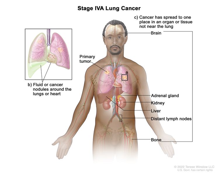
Cáncer de pulmón en estadio IVA. El tumor puede ser de cualquier tamaño y el cáncer puede haberse extendido a los ganglios linfáticos. Se da una o más de las siguientes condiciones: (a) hay uno o más tumores en el pulmón que no tiene el tumor primario; y/o (b) se encuentra cáncer en el líquido que rodea los pulmones o el corazón, o hay nódulos cancerosos en el revestimiento que rodea los pulmones o el saco que rodea el corazón; y/o (c) el cáncer se ha extendido a un lugar en un órgano o tejido que no está cerca del pulmón, en particular el cerebro, la glándula suprarrenal, el riñón, el hígado o los huesos, o a un ganglio linfático que no está cerca del pulmón.
El tumor puede ser de cualquier tamaño y el cáncer puede haberse diseminado a los ganglios linfáticos . Se encuentra uno o más de los siguientes:
- Hay uno o más tumores en el pulmón que no tienen el tumor primario .
- El cáncer se encuentra en el revestimiento que rodea los pulmones o en el saco que rodea el corazón.
- El cáncer se encuentra en el líquido que rodea los pulmones o el corazón.
- El cáncer se ha extendido a un órgano que no está cerca del pulmón, como el cerebro, el hígado, la glándula suprarrenal, el riñón, el hueso o un ganglio linfático que no está cerca del pulmón.
- Estadio IVB:
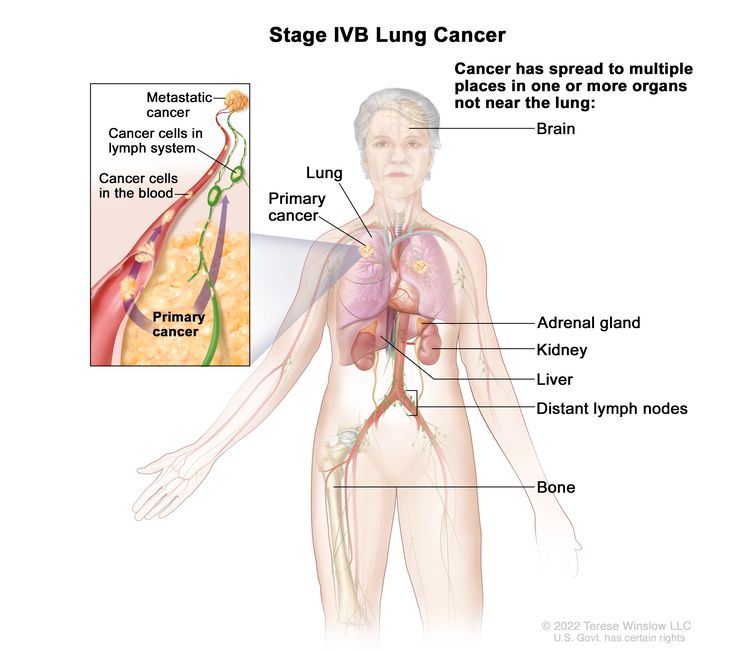
Cáncer de pulmón en estadio IVB. El cáncer se ha diseminado a múltiples lugares en uno o más órganos que no están cerca del pulmón, como el cerebro, la glándula suprarrenal, el riñón, el hígado, los ganglios linfáticos distantes o el hueso.
El cáncer se ha extendido a múltiples lugares en uno o más órganos que no están cerca del pulmón.
El cáncer de pulmón de células no pequeñas puede recidivar (regresar) después de haber sido tratado.
El cáncer de pulmón de células no pequeñas recurrente es aquel que ha reaparecido después de haber sido tratado. Si el cáncer de pulmón de células no pequeñas reaparece, puede hacerlo en el cerebro, el pulmón, el tórax o en otras partes del cuerpo. Se realizarán pruebas para ayudar a determinar dónde ha reaparecido el cáncer. El tipo de tratamiento para el cáncer de pulmón de células no pequeñas dependerá de dónde haya reaparecido.
Learn more in Recurrent Cancer: When Cancer Comes Back.
Treatment Option Overview
Puntos clave
- Existen diferentes tipos de tratamiento para personas con cáncer de pulmón de células no pequeñas.
- The following types of treatment are used:
- Cirugía
- Radioterapia
- Quimioterapia
- Terapia dirigida
- Inmunoterapia
- Terapia con láser
- Terapia fotodinámica (TFD)
- Cryosurgery
- Electrocauterización
- New types of treatment are being tested in clinical trials.
- El tratamiento para el cáncer de pulmón de células no pequeñas puede causar efectos secundarios.
- Es posible que se necesiten cuidados de seguimiento.
Existen diferentes tipos de tratamiento para personas con cáncer de pulmón de células no pequeñas.
Existen distintos tipos de tratamientos disponibles para las personas con cáncer de pulmón de células no pequeñas. Usted y su equipo de atención médica del cáncer trabajarán juntos para definir el plan de tratamiento más adecuado, que puede incluir más de una opción terapéutica. Se tomarán en cuenta diversos factores, como el estadio y el grado del cáncer, su estado de salud general y sus preferencias. El plan contendrá información sobre su enfermedad, los objetivos del tratamiento, las alternativas disponibles, los posibles efectos secundarios y la duración estimada del proceso.
Hablar con tu equipo de atención oncológica antes de que comience el tratamiento sobre qué esperar te será de gran ayuda. Es importante que sepas qué debes hacer antes de que empiece el tratamiento, cómo te sentirás durante el mismo y qué tipo de ayuda necesitarás. Para obtener más información, visita la sección Preguntas para hacerle a tu médico sobre el tratamiento .
The following types of treatment are used:
Cirugía
Para tratar el cáncer de pulmón se utilizan cuatro tipos de cirugía:
- La resección en cuña es una cirugía para extirpar un tumor y parte del tejido normal que lo rodea. Cuando se toma una cantidad de tejido ligeramente mayor, se denomina resección segmentaria.
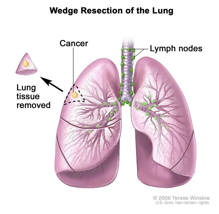
Resección en cuña del pulmón. Se extirpa parte del lóbulo del pulmón que contiene el cáncer y una pequeña cantidad de tejido sano a su alrededor.
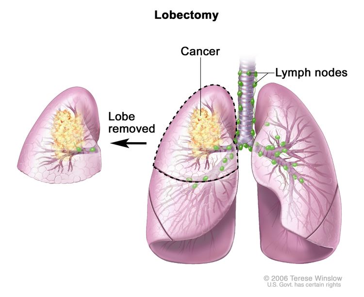
- La lobectomía es una cirugía para extirpar un lóbulo completo (sección) del pulmón.
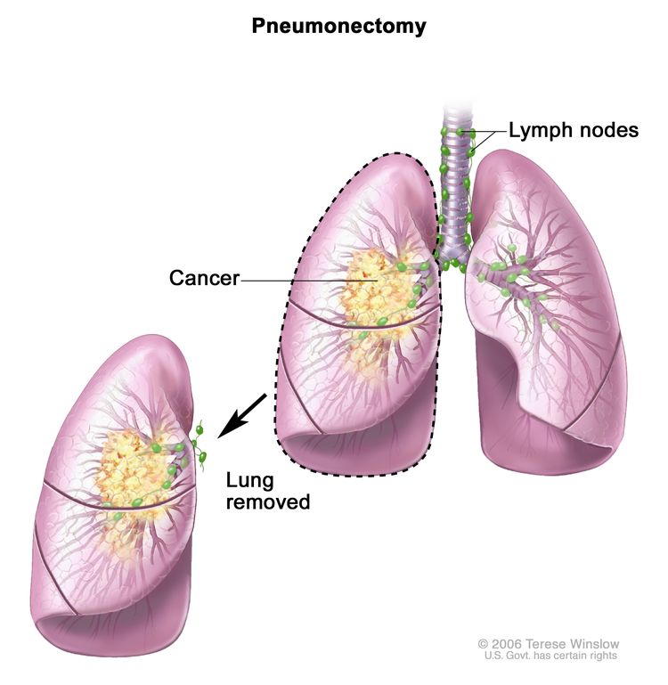
- La neumonectomía es una cirugía para extirpar un pulmón entero.
- La resección del manguito es una cirugía para extirpar parte del bronquio.
Después de que el médico extirpa todo el cáncer que se puede ver en el momento de la cirugía, algunas personas pueden recibir quimioterapia o radioterapia para eliminar cualquier célula cancerosa restante. El tratamiento que se administra después de la cirugía para reducir el riesgo de que el cáncer regrese se llama terapia adyuvante.
Radioterapia
La radioterapia es un tratamiento contra el cáncer que utiliza rayos X de alta energía u otros tipos de radiación para matar las células cancerosas o evitar que crezcan. Hay dos tipos de radioterapia:
- Radioterapia externa Utiliza una máquina externa al cuerpo para enviar radiación hacia la zona con cáncer. Ciertas formas de administrar radioterapia externa pueden ayudar a evitar que la radiación dañe el tejido sano cercano:
- La radioterapia corporal estereotáctica utiliza un equipo especial para garantizar que el paciente permanezca en la misma posición durante cada sesión de radiación. Una vez al día, durante varios días, un aparato de radiación aplica una dosis de radiación mayor de lo habitual directamente al tumor. Al mantenerlo en la misma posición durante cada sesión, se reduce el daño al tejido sano circundante. Este procedimiento también se denomina radioterapia estereotáctica de haz externo y radioterapia estereotáxica.
- La radiocirugía estereotáctica se utiliza para tratar el cáncer de pulmón que se ha propagado al cerebro. Se coloca un marco rígido en el cráneo para mantener la cabeza inmóvil durante el tratamiento con radiación. Una máquina dirige una sola dosis grande de radiación directamente al tumor en el cerebro. Este procedimiento no implica cirugía. También se denomina radiocirugía estereotáctica, radiocirugía y cirugía con radiación.
- La radioterapia interna utiliza una sustancia radiactiva sellada en agujas, semillas, cables o catéteres que se colocan directamente en el cáncer o cerca de él.
Para los tumores en las vías respiratorias, la radiación se administra directamente al tumor a través de un endoscopio.
La forma en que se administra la radioterapia depende del tipo y estadio del cáncer que se está tratando. También depende de dónde se encuentre el cáncer. La radioterapia externa e interna se usa para tratar el cáncer de pulmón de células no pequeñas.
Quimioterapia
La quimioterapia (también llamada quimio) utiliza fármacos para detener el crecimiento de las células cancerosas, ya sea matándolas o impidiendo que se dividan.
La quimioterapia para el cáncer de pulmón de células no pequeñas suele ser sistémica, es decir, se inyecta en una vena o se administra por vía oral. Cuando se administra de esta manera, los medicamentos ingresan al torrente sanguíneo para llegar a las células cancerosas en todo el cuerpo.
Los medicamentos de quimioterapia utilizados para tratar el cáncer de pulmón de células no pequeñas pueden incluir:
- carboplatin
- Cisplatino
- docetaxel
- doxorubicin
- Etopósido
- gemcitabine
- paclitaxel
- Pemetrexed
- Vinorelbina
Se pueden utilizar combinaciones de estos fármacos de quimioterapia. También se pueden utilizar otros fármacos de quimioterapia no mencionados aquí.
La quimioterapia también puede combinarse con otros tipos de tratamiento. Por ejemplo, puede combinarse con radioterapia o fármacos de inmunoterapia .
Para obtener más información sobre cómo funciona la quimioterapia, cómo se administra, los efectos secundarios comunes y mucho más, visite Quimioterapia para tratar el cáncer y Quimioterapia y usted: Apoyo para personas con cáncer .
Terapia dirigida
Targeted therapy uses drugs or other substances to identify and attack specific cancer cells. Your doctor may suggest biomarker tests to help predict your response to certain targeted therapy drugs. Learn more about Biomarker Testing for Cancer Treatment.
Entre las terapias dirigidas utilizadas para tratar el cáncer de pulmón de células no pequeñas están:
- Adagrasib
- Afatinib
- Alectinib
- Amivantamab
- Bevacizumab
- Brigatinib
- capmatinib
- ceritinib
- Cetuximab
- crizotinib
- dabrafenib
- Dacomitinib
- entrectinib
- erlotinib
- Everolimus
- gefitinib
- larotrectinib
- Lazertinib
- lorlatinib
- Necitumumab
- Osimertinib
- Pralsetinib
- Ramucirumab
- Repotrectinib
- selpercatinib
- Sotorasib
- tepotinib
- trametinib
Obtenga más información en Terapia dirigida para tratar el cáncer.
Inmunoterapia
Immunotherapy helps a person's immune system fight cancer. Your doctor may suggest biomarker tests to help predict your response to certain immunotherapy drugs. Learn more about Biomarker Testing for Cancer Treatment.
Los medicamentos de inmunoterapia utilizados para tratar el cáncer de pulmón de células no pequeñas son:
Obtenga más información sobre la inmunoterapia para el tratamiento del cáncer .
Terapia con láser
La terapia láser es un tratamiento contra el cáncer que utiliza un rayo láser (un haz estrecho de luz intensa) para destruir las células cancerosas.
Más información sobre los láseres para tratar el cáncer.
Terapia fotodinámica (TFD)
La terapia fotodinámica (TFD) es un tratamiento contra el cáncer que utiliza un fármaco y un tipo específico de luz láser para destruir las células cancerosas. Se inyecta en una vena un fármaco que permanece inactivo hasta que se expone a la luz. Este fármaco se acumula en mayor cantidad en las células cancerosas que en las células sanas. Posteriormente, se utilizan tubos de fibra óptica para dirigir la luz láser hacia las células cancerosas, donde el fármaco se activa y las destruye. La terapia fotodinámica causa poco daño al tejido sano. Se utiliza principalmente para tratar tumores en la piel o justo debajo de ella, o en el revestimiento de los órganos internos. Cuando el tumor se encuentra en las vías respiratorias, la TFD se administra directamente al tumor mediante un endoscopio.
Más información sobre la terapia fotodinámica para tratar el cáncer.
Cryosurgery
La criocirugía es un tratamiento que utiliza un instrumento para congelar y destruir tejido anómalo, como el carcinoma in situ. Este tipo de tratamiento también se denomina crioterapia. Para los tumores en las vías respiratorias, la criocirugía se realiza mediante un endoscopio.
Más información sobre la criocirugía para tratar el cáncer.
Electrocauterización
El electrocauterio es un tratamiento que utiliza una sonda o aguja calentada por una corriente eléctrica para destruir el tejido anómalo. En el caso de los tumores de las vías respiratorias, el electrocauterio se realiza a través de un endoscopio.
New types of treatment are being tested in clinical trials.
For some people, joining a clinical trial may be an option. There are different types of clinical trials for people with cancer. For example, a treatment trial tests new treatments or new ways of using current treatments. Supportive care and palliative care trials look at ways to improve quality of life, especially for those who have side effects from cancer and its treatment.
You can use the clinical trial search to find NCI-supported cancer clinical trials accepting participants. The search allows you to filter trials based on the type of cancer, your age, and where the trials are being done. Clinical trials supported by other organizations can be found on the ClinicalTrials.gov website.
Para más información sobre ensayos clínicos, cómo encontrarlos y participar en uno de ellos, visite la web Información sobre estudios clínicos para pacientes y cuidadores.
El tratamiento para el cáncer de pulmón de células no pequeñas puede causar efectos secundarios.
For information about side effects caused by treatment for cancer, visit our Side Effects page.
Es posible que se necesiten cuidados de seguimiento.
A medida que avanza el tratamiento, se le realizarán pruebas o controles de seguimiento. Es posible que se repitan algunas pruebas para diagnosticar o estadificar el cáncer con el fin de evaluar cómo está funcionando el tratamiento. Las decisiones sobre si continuar, modificar o suspender el tratamiento pueden basarse en los resultados de estas pruebas.
Algunas pruebas seguirán realizándose de manera periódica después de terminar el tratamiento. Los resultados pueden indicar si su afección ha cambiado o si el cáncer ha redicivado (regresado).
Tratamiento del cáncer de pulmón de células no pequeñas oculto
El tratamiento del cáncer de pulmón de células no pequeñas oculto depende del estadio de la enfermedad. Los tumores ocultos suelen detectarse en una fase temprana (el tumor se encuentra únicamente en el pulmón) y, en ocasiones, pueden curarse mediante cirugía.
Puede utilizar la búsqueda de ensayos clínicos y encontrar ensayos clínicos sobre cáncer patrocinados por el NCI que acepten participantes. La búsqueda le permite filtrar los ensayos según el tipo de cáncer, la edad y el lugar donde se realizan los ensayos. También encontrará información general sobre los ensayos clínicos.
Treatment of Stage 0 (carcinoma in situ)
El tratamiento de la etapa 0 puede incluir:
- Cirugía (resección en cuña o resección segmentaria)
- terapia fotodinámica, electrocauterización, criocirugía o cirugía láser para tumores en o cerca de los bronquios.
Learn more about these treatments in the Treatment Option Overview.
Puede utilizar la búsqueda de ensayos clínicos y encontrar ensayos clínicos sobre cáncer patrocinados por el NCI que acepten participantes. La búsqueda le permite filtrar los ensayos según el tipo de cáncer, la edad y el lugar donde se realizan los ensayos. También encontrará información general sobre los ensayos clínicos.
Tratamiento del cáncer de pulmón de células no pequeñas en estadio I
El tratamiento del cáncer de pulmón de células no pequeñas en estadio IA y del cáncer de pulmón de células no pequeñas en estadio IB puede implicar:
- Cirugía (resección en cuña, resección segmentaria, resección en manguito o lobectomía)
- Cirugía seguida de terapia dirigida
- Cirugía seguida de quimioterapia e inmunoterapia
- radioterapia externa, incluida la radioterapia corporal estereotáctica para personas que no pueden someterse a cirugía o que eligen no someterse a ella.
Más información sobre estos tratamientos y encuentre una lista de medicamentos de quimioterapia, terapia dirigida e inmunoterapia para el cáncer de pulmón en la Descripción general de opciones de tratamiento.
Puede utilizar la búsqueda de ensayos clínicos y encontrar ensayos clínicos sobre cáncer patrocinados por el NCI que acepten participantes. La búsqueda le permite filtrar los ensayos según el tipo de cáncer, la edad y el lugar donde se realizan los ensayos. También encontrará información general sobre los ensayos clínicos.
Tratamiento del cáncer de pulmón de células no pequeñas en estadio II
El tratamiento del cáncer de pulmón de células no pequeñas en estadio IIA y del cáncer de pulmón de células no pequeñas en estadio IIB puede ser:
- Cirugía (resección en cuña, resección segmentaria, resección en manguito, lobectomía o neumonectomía)
- Cirugía seguida de quimioterapia
- Cirugía seguida de terapia dirigida
- Cirugía seguida de quimioterapia e inmunoterapia
- Cirugía seguida de inmunoterapia
- cirugía seguida de radioterapia
- Quimioterapia seguida de cirugía
- Inmunoterapia y quimioterapia seguidas de cirugía
- Inmunoterapia y quimioterapia seguidas de cirugía y más inmunoterapia
- Radioterapia externa para personas que no pueden someterse a cirugía
Más información sobre estos tratamientos y encuentre una lista de medicamentos de quimioterapia, terapia dirigida e inmunoterapia para el cáncer de pulmón en la Descripción general de opciones de tratamiento.
Puede utilizar la búsqueda de ensayos clínicos y encontrar ensayos clínicos sobre cáncer patrocinados por el NCI que acepten participantes. La búsqueda le permite filtrar los ensayos según el tipo de cáncer, la edad y el lugar donde se realizan los ensayos. También encontrará información general sobre los ensayos clínicos.
Tratamiento del cáncer de pulmón de células no pequeñas en estadio IIIA
El tratamiento del cáncer de pulmón de células no pequeñas en estadio IIIA que se puede extirpar mediante cirugía puede incluir:
- Quimioterapia seguida de cirugía
- Quimioterapia y radioterapia seguidas de cirugía
- Inmunoterapia y quimioterapia seguidas de cirugía
- Inmunoterapia y quimioterapia seguidas de cirugía y más inmunoterapia
- Cirugía seguida de quimioterapia
- Cirugía seguida de terapia dirigida
- Cirugía seguida de quimioterapia e inmunoterapia
- Cirugía seguida de inmunoterapia
- Cirugía seguida de quimioterapia y radioterapia.
- Cirugía seguida de radioterapia
El tratamiento del cáncer de pulmón de células no pequeñas en estadio IIIA, cuando no se puede extirpar con cirugía, puede incluir:
- Quimioterapia y radioterapia
- Quimioterapia y radioterapia seguidas de inmunoterapia
- Radioterapia externa sola
- Radioterapia interna o cirugía láser como tratamiento paliativo para aliviar los síntomas y mejorar la calidad de vida
Más información sobre atención de apoyo para signos y síntomas como tos, dificultad para respirar y dolor en el pecho en las secciones Síndromes cardiopulmonares y El dolor y el cáncer.
El cáncer de pulmón de células no pequeñas del surco superior, conocido como tumor de Pancoast, se origina en la parte superior del pulmón y puede propagarse a tejidos cercanos como la pared torácica, los grandes vasos sanguíneos y la columna vertebral. El tratamiento de los tumores de Pancoast puede incluir:
- Cirugía
- Quimioterapia y radioterapia seguidas de cirugía
- Radioterapia sola
Algunos tumores pulmonares de células no pequeñas en estadio IIIA que han invadido la pared torácica pueden extirparse por completo. El tratamiento para estos tumores puede contemplar:
- Cirugía
- Cirugía y radioterapia
- Radioterapia sola
- Quimioterapia combinada con radioterapia y/o cirugía
Más información sobre estos tratamientos y encuentre una lista de medicamentos de quimioterapia para el cáncer de pulmón en la Descripción general de opciones de tratamiento .
Puede utilizar la búsqueda de ensayos clínicos y encontrar ensayos clínicos sobre cáncer patrocinados por el NCI que acepten participantes. La búsqueda le permite filtrar los ensayos según el tipo de cáncer, la edad y el lugar donde se realizan los ensayos. También encontrará información general sobre los ensayos clínicos.
Tratamiento del cáncer de pulmón de células no pequeñas en estadio IIIB y estadio IIIC
El tratamiento del cáncer de pulmón de células no pequeñas en estadio IIIB y del cáncer de pulmón de células no pequeñas en estadio IIIC puede consistir en:
- Quimioterapia seguida de radioterapia externa
- Quimioterapia con radioterapia
- Inmunoterapia y quimioterapia seguidas de cirugía y más inmunoterapia
- Inmunoterapia antes o después de la quimioterapia y la radioterapia
- Terapia dirigida antes o después de la quimioterapia y la radioterapia
- Radioterapia externa únicamente para personas que no pueden recibir quimioterapia
- Radioterapia externa como terapia paliativa para aliviar los síntomas y mejorar la calidad de vida
- Terapia con láser y/o radioterapia interna para aliviar los síntomas y mejorar la calidad de vida
Más información sobre estos tratamientos y encuentre una lista de medicamentos de quimioterapia, terapia dirigida e inmunoterapia para el cáncer de pulmón en la Descripción general de opciones de tratamiento.
Más información sobre atención de apoyo para signos y síntomas como tos, dificultad para respirar y dolor en el pecho en las secciones Síndromes cardiopulmonares y El dolor y el cáncer.
Puede utilizar la búsqueda de ensayos clínicos y encontrar ensayos clínicos sobre cáncer patrocinados por el NCI que acepten participantes. La búsqueda le permite filtrar los ensayos según el tipo de cáncer, la edad y el lugar donde se realizan los ensayos. También encontrará información general sobre los ensayos clínicos.
Tratamiento del cáncer de pulmón de células no pequeñas en estadio IV, recidivante y recurrente, que se ha diagnosticado recientemente
Tratamiento del cáncer de pulmón de células no pequeñas en estadio IV, recidivante y recurrente puede incluir:
- Uno o más medicamentos de quimioterapia con o sin terapia dirigida
- Quimioterapia combinada seguida de más quimioterapia como terapia de mantenimiento para ayudar a evitar que el cáncer avance
- Terapia dirigida
- Uno o más medicamentos de inmunoterapia
Más información sobre estos tratamientos y encuentre una lista de medicamentos de quimioterapia, terapia dirigida e inmunoterapia para el cáncer de pulmón en la Descripción general de opciones de tratamiento.
Puede utilizar la búsqueda de ensayos clínicos y encontrar ensayos clínicos sobre cáncer patrocinados por el NCI que acepten participantes. La búsqueda le permite filtrar los ensayos según el tipo de cáncer, la edad y el lugar donde se realizan los ensayos. También encontrará información general sobre los ensayos clínicos.
Tratamiento del cáncer de pulmón de células no pequeñas en estadio IV progresivo, recidivante y recurrente
El tratamiento del cáncer de pulmón de células no pequeñas en estadio IV progresivo, recidivante y recurrente puede incluir:
- Quimioterapia
- terapia dirigida con o sin quimioterapia
- Inmunoterapia
Más información sobre estos tratamientos y encuentre una lista de medicamentos de quimioterapia, terapia dirigida e inmunoterapia para el cáncer de pulmón en la Descripción general de opciones de tratamiento.
Puede utilizar la búsqueda de ensayos clínicos y encontrar ensayos clínicos sobre cáncer patrocinados por el NCI que acepten participantes. La búsqueda le permite filtrar los ensayos según el tipo de cáncer, la edad y el lugar donde se realizan los ensayos. También encontrará información general sobre los ensayos clínicos.
Más información sobre el cáncer de pulmón de células no pequeñas
Para obtener más información del National Cancer Institute sobre el cáncer de pulmón de células no pequeñas, visite:
- Página principal sobre el cáncer de pulmón
- Lung Cancer Prevention
- Detección del cáncer de pulmón
- Medicamentos aprobados para el cáncer de pulmón de células no pequeñas
- Tabaco (incluye ayuda para dejarlo)
- Humo de tabaco en el ambiente y el cáncer
For general cancer information and other resources from the National Cancer Institute, visit:
Sobre este resumen del PDQ
Acerca del PDQ
El Physician Data Query (PDQ) es la base de datos integral sobre el cáncer del National Cancer Institute (NCI). La base de datos del PDQ contiene resúmenes con la última información publicada sobre prevención, detección, genética, tratamiento, atención médica de apoyo y medicina complementaria y alternativa relacionada con el cáncer. La mayoría de los resúmenes se presentan en dos versiones. Las versiones para profesionales de la salud contienen información detallada escrita en lenguaje técnico. Las versiones para pacientes están escritas en un lenguaje fácil de entender y no tan técnico. Ambas versiones contienen información precisa y actualizada sobre el cáncer. La mayoría de las versiones también están disponibles en español.
El PDQ es un servicio del NCI. El NCI es parte de los Institutos Nacionales de Salud (NIH), que son el centro de investigación biomédica del Gobierno federal. Los resúmenes del PDQ se basan en una revisión independiente de la literatura médica. No son declaraciones de políticas del NCI ni de los NIH.
Propósito de este resumen
Este resumen del PDQ sobre el cáncer contiene información actualizada del tratamiento del cáncer de pulmón de células no pequeñas. El propósito es informar y ayudar a los pacientes, sus familiares y cuidadores. No da pautas ni recomendaciones formales para tomar decisiones relacionadas con la atención médica.
Revisores y actualizaciones
Los comités editoriales escriben los resúmenes de información sobre el cáncer del PDQ y los mantienen actualizados. Estos comités están formados por equipos de especialistas en el tratamiento del cáncer y otras especialidades relacionadas con esta enfermedad. Los resúmenes se revisan periódicamente y se modifican cuando hay información nueva. La fecha de actualización al pie de cada resumen indica cuándo se realizó el cambio más reciente.
The information in this patient summary was taken from the health professional version, which is reviewed regularly and updated as needed, by the PDQ Adult Treatment Editorial Board.
Información sobre ensayos clínicos
Un ensayo clínico es un estudio para responder a una pregunta científica como, por ejemplo, si un tratamiento es mejor que otro. Los ensayos se basan en estudios anteriores y en lo aprendido en el laboratorio. Cada ensayo responde a determinadas preguntas científicas que permiten encontrar nuevas y mejores formas de ayudar a los pacientes con cáncer. Durante los ensayos clínicos de tratamiento, se recopila información sobre los efectos de un nuevo tratamiento y su eficacia. Si un ensayo clínico demuestra que un nuevo tratamiento es mejor que uno que se utiliza actualmente, el nuevo tratamiento puede convertirse en “estándar”. Los pacientes pueden valorar la posibilidad de participar en un ensayo clínico. Algunos ensayos clínicos solo están abiertos a pacientes que no hayan iniciado el tratamiento.
Los ensayos clínicos se pueden encontrar en línea en el sitio web del NCI. Para obtener más información, llame al Servicio de Información sobre el Cáncer (CIS, por sus siglas en inglés), el centro de contacto del NCI, al 1-800-4-CANCER (1-800-422-6237).
Permiso de uso de este resumen
Physician Data Query (PDQ) es una marca registrada. Se autoriza el libre uso del contenido de los documentos del PDQ como texto. Sin embargo, no se podrá identificar como un resumen de información sobre cáncer del PDQ del NCI, salvo que se reproduzca en su totalidad y se actualice con regularidad. Por otra parte, se permite que los autores incluyan una oración como “en el resumen del PDQ del NCI sobre la prevención del cáncer de mama se describen, de manera concisa, los siguientes riesgos: [incluir fragmento del resumen]”.
La forma recomendada para citar este resumen del PDQ es:
Comité editorial del PDQ® sobre el tratamiento para adultos. Tratamiento del cáncer de pulmón de células no pequeñas (PDQ). Bethesda, MD: National Cancer Institute. Actualizado el [DD/MM/AAAA]
Las imágenes de este resumen se utilizan con el permiso del autor, artista y/o editorial para uso exclusivo en los resúmenes del PDQ. Si desea usar una imagen de un resumen del PDQ sin incluir el resumen completo, debe obtener autorización del propietario. El National Cancer Institute no puede otorgar dicho permiso. Para obtener más información sobre el uso de las imágenes de este resumen o de otras ilustraciones relacionadas con el cáncer, consulte Visuals Online, una colección de más de 3,000 imágenes científicas.
Descargo de responsabilidad
La información de estos resúmenes no debe utilizarse para tomar decisiones sobre reembolsos de seguros. Puede encontrar más información sobre la cobertura de seguros en Cancer.gov en el sitio Manejo de la atención del cáncer.
Contáctenos
Puede encontrar más información sobre cómo contactarnos o recibir ayuda en el sitio web Cancer.gov en la página Comuníquese con el NCI. También puede enviar sus preguntas a Cancer.gov en el apartado Escríbanos del sitio web.
Actualizado:
URL de origen: https://www.cancer.gov/node/4162/syndication
Agencia de origen: National Cancer Institute (NCI)
Captured Date: 2013-09-14 09:02:03.0