


Tratamiento del cáncer de vagina
Acceda a una atención excepcional para el cáncer vaginal y otros tipos de cánceres ginecológicos en Montefiore Einstein Comprehensive Cancer Center. Nuestro equipo multidisciplinario tiene una amplia experiencia en el tratamiento de este cáncer. Somos uno de los primeros centros del cáncer designados por el NCI y, como tales, llevamos más de 50 años siendo líderes en la investigación, el diagnóstico y el tratamiento de más de 200 tipos de cáncer.
Estamos a la vanguardia en el tratamiento y la investigación del cáncer vaginal y otros cánceres ginecológicos. Ofrecemos planes de tratamiento integrales y personalizados según las necesidades de las pacientes. En Montefiore Einstein Comprehensive Cancer Center, ofrecemos acceso a atención de vanguardia e innovadora, basada en las últimas investigaciones sobre el cáncer.
Cuando necesite atención médica para el cáncer de vagina, confíe en nuestros proveedores, quienes se dedican con pasión a combatir el cáncer y a atender todas sus necesidades de salud.

El Montefiore Einstein Comprehensive Cancer Center, designado como centro integral del cáncer por el National Cancer Institute (NCI), apoya la misión y las normas del NCI. La siguiente información sobre los tipos de cáncer, prevención y tratamientos ha sido facilitada por el NCI.
Tratamiento del cáncer de vagina (PDQ®): versión para pacientes
Información general sobre el cáncer de vagina
Puntos clave
- El cáncer vaginal es una enfermedad en la que se forman células malignas (cancerosas) en la vagina.
- La edad avanzada y la infección por VPH son factores de riesgo para el cáncer de vagina.
- Los signos y síntomas del cáncer vaginal incluyen dolor o sangrado anómalo en la vagina.
- Para diagnosticar el cáncer de vagina, se utilizan pruebas que examinan la vagina y otros órganos de la pelvis.
- Certain factors affect prognosis (chance of recovery) and treatment options.
El cáncer vaginal es una enfermedad en la que se forman células malignas (cancerosas) en la vagina.
La vagina es el conducto que va desde el cuello uterino (la abertura del útero) al exterior del cuerpo. En el parto, el bebé sale del cuerpo a través de la vagina (también llamada canal de parto).
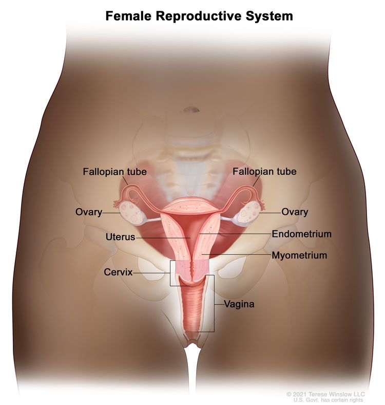
Anatomía del aparato reproductor femenino. Entre los órganos del aparato reproductor femenino están el útero, los ovarios, las trompas de Falopio, el cuello uterino y la vagina. El útero tiene una capa muscular externa, llamada miometrio, y una capa de tejido interno que lo reviste, llamada endometrio.
El cáncer vaginal no es común. Existen dos tipos principales de cáncer vaginal:
- Carcinoma escamocelular: cáncer que se forma en las células delgadas y planas que recubren el interior de la vagina. El cáncer vaginal de células escamosas se propaga lentamente y suele mantenerse cerca de la vagina, pero puede extenderse a los pulmones, el hígado o los huesos. Es el tipo más común de cáncer vaginal.
- Adenocarcinoma: cáncer que comienza en las células glandulares. Las células glandulares del revestimiento de la vagina producen y liberan líquidos como las secreciones mucosas. El adenocarcinoma tiene más probabilidades que el carcinoma escamocelular de propagarse a los pulmones y los ganglios linfáticos. Un tipo raro de adenocarcinoma (adenocarcinoma de células claras) está relacionado con la exposición al dietilestilbestrol (DES) antes del nacimiento. Es más frecuente que los adenocarcinomas que no están relacionados con la exposición al DES aparezcan en las mujeres después de la menopausia.
La edad avanzada y la infección por VPH son factores de riesgo para el cáncer de vagina.
Todo lo que aumenta las posibilidades de que una persona tenga una enfermedad se llama factor de riesgo. No todas las personas con uno o más de estos factores de riesgo desarrollarán cáncer de vagina, y algunas personas sin factores de riesgo conocidos presentarán la enfermedad. Consulte a su médico si cree que está en riesgo. Los factores de riesgo del cáncer de vagina son los siguientes:
- Tener 60 años o más
- Tener una infección por el virus del papiloma humano (VPH), que a veces se relaciona con el carcinoma escamocelular de la vagina.
- Haber estado expuesta al DES en el útero materno. En la década de 1950, algunas mujeres embarazadas recibieron DES, un medicamento para prevenir abortos espontáneos (nacimientos prematuros de fetos que no sobreviven). Esto se relaciona con una forma rara de cáncer vaginal llamado adenocarcinoma de células claras. Las tasas de incidencia de esta enfermedad tocaron su máximo en la década de 1970, pero, en la actualidad, es extremadamente rara.
- Haberse realizado una histerectomía por tumores benignos (no cancerosos) o por cáncer
Los signos y síntomas del cáncer vaginal incluyen dolor o sangrado anómalo en la vagina.
A menudo, el cáncer de vagina no causa signos o síntomas tempranos. Es posible que se lo detecte durante un examen pélvico y una prueba de Papanicolao de rutina. Los signos y síntomas pueden deberse al cáncer de vagina o a otras afecciones. Consulte con su médico si tiene alguno de los siguientes signos y síntomas:
- Sangrados o secreciones no relacionados con el periodo menstrual
- Dolor durante las relaciones sexuales
- Dolor en la zona pélvica
- Un bulto en la vagina
- Dolor al orinar
- Constipación.
Para diagnosticar el cáncer de vagina, se utilizan pruebas que examinan la vagina y otros órganos de la pelvis.
In addition to asking about your personal and family health history and doing a physical exam, your doctor may perform the following tests and procedures:
- Examen pélvico: un examen de la vagina, cuello uterino, útero, trompas de Falopio, ovarios, and recto. Un espéculo se introduce en la vagina y el médico o enfermero examina la vagina y el cuello uterino en busca de signos de enfermedad. Por lo general, se realiza una prueba de Papanicolaou del cuello uterino. El médico o enfermero también inserta uno o dos lubricados, dedos enguantados de una mano en la vagina y coloca la otra mano sobre la parte inferior del abdomen para palpar el tamaño, la forma y la posición del útero y los ovarios. El médico o enfermero también inserta un dedo enguantado y lubricado en el recto para palpar bultos o anómalas zonas.
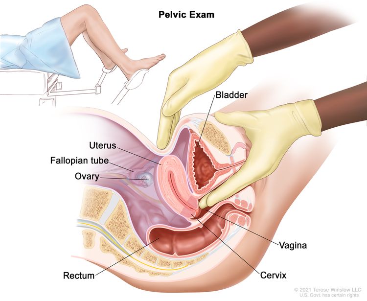
Examen pélvico. Un médico o enfermero introduce uno o dos dedos de una mano, con guantes y lubricados, en la vagina, mientras presiona la parte inferior del abdomen con la otra mano. Esto se hace para palpar el tamaño, la forma y la posición del útero y los ovarios. También se examinan la vagina, el cuello uterino, las trompas de Falopio y el recto.
- Prueba de Papanicolaou: procedimiento para recolectar células de la superficie del cuello uterino y la vagina. Se utiliza un trozo de algodón, un cepillo o una espátula pequeña de madera para hacer un raspado suave de las células del cuello uterino y la vagina. Las células se observan con microscope para saber si son anormales. Este procedimiento también se llama prueba de Papanicolaou.
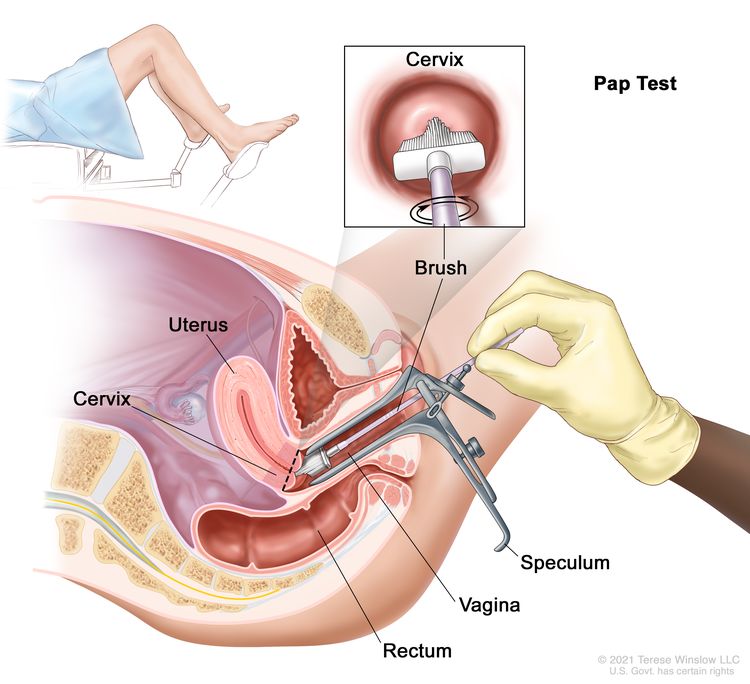
Prueba de Pap. Se introduce un espéculo en la vagina para ensancharla. A continuación, se introduce un cepillo para recolectar células del cuello uterino. Las células se examinan con microscopio para detectar signos de enfermedad.
- Prueba del virus del papiloma humano (VPH): prueba de laboratorio a través de la que se detecta el ADN o el ARN de ciertos tipos de VPH. Se recolectan células del cuello uterino y se analiza su ADN o ARN para determinar si una infección es consecuencia de un tipo de VPH relacionado con el cáncer de cuello uterino. Para esta prueba, se puede usar la muestra de células extraída durante una prueba de Pap. Esta prueba también se hace cuando los resultados de una prueba de Pap indican que hay ciertas células anómalas en el cuello del útero.
- colposcopia: procedimiento para el que se utiliza un colposcopio (un instrumento con aumento y luz) para examinar la vagina y el cuello uterino en busca de áreas anómalas. Se pueden tomar muestras de tejido con una cureta (instrumento con forma de cuchara) o un cepillo y examinarlas bajo un microscopio para detectar signos de enfermedad.
- La biopsia: Extracción de células o tejidos de la vagina y el cuello uterino para que un patólogo los examine al microscopio y detecte signos de cáncer. Si una prueba de Papanicolaou muestra células anormales en la vagina, se puede realizar una biopsia durante una colposcopia.
Certain factors affect prognosis (chance of recovery) and treatment options.
El pronóstico depende de los siguientes factores:
- El estadio del cáncer (si afecta solo la vagina o se ha diseminado a otras áreas)
- El tamaño del tumor
- El grado de las células tumorales (qué tan diferentes se ven de las células normales bajo un microscopio)
- La ubicación del cáncer en la vagina
- La presencia de signos o síntomas al momento del diagnóstico
- Si el cáncer acaba de ser diagnosticado o ha recidivado (regresado)
Las opciones de tratamiento dependen de los siguientes factores:
- El estadio y el tamaño del cáncer
- Si el cáncer está cerca de otros órganos que puedan resultar dañados por el tratamiento.
- Si el tumor está formado por células escamosas o es un adenocarcinoma.
- Si la paciente tiene útero o se ha realizado una histerectomía.
- Si la paciente ha recibido radioterapia en la pelvis en el pasado.
Estadios del cáncer de vagina
Puntos clave
- Después de diagnosticar un cáncer de vagina, se realizan pruebas para determinar si las células cancerosas se han diseminado dentro de la vagina o hacia otras partes del cuerpo.
- There are three ways that cancer spreads in the body.
- El cáncer puede extenderse desde donde comenzó a otras partes del cuerpo.
- En la neoplasia intraepitelial vaginal (NIV) se encuentran células anómalas en el tejido que reviste el interior de la vagina.
- Para clasificar el cáncer de vagina se utilizan los siguientes estadios:
- Estadio I
- Estadio II
- Estadio III
- Estadio IV
- El cáncer de vagina puede recidivar (regresar) después de haber sido tratado.
Después de diagnosticar un cáncer de vagina, se realizan pruebas para determinar si las células cancerosas se han diseminado dentro de la vagina o hacia otras partes del cuerpo.
El proceso que se utiliza para determinar si el cáncer se ha propagado dentro de la vagina o a otras partes del cuerpo se denomina estadificación. La información obtenida del proceso de estadificación determina el estadio de la enfermedad. Es importante conocer el estadio para planificar el tratamiento. Es posible que se usen los siguientes procedimientos en el proceso de estadificación:
- Radiografía del tórax: radiografía de los órganos y huesos del interior del tórax. Los rayos X son un tipo de haz de energía que puede atravesar el cuerpo, plasmarse en una película y generar una imagen de áreas del interior del cuerpo.
- Tomografía computarizada (TC): procedimiento que genera una serie de imágenes detalladas de áreas internas del cuerpo como el abdomen o la pelvis, desde diferentes ángulos. Las imágenes se crean con una computadora conectada a una máquina de rayos X. A veces, se inyecta un tinte en una vena o se ingiere para ayudar a que los órganos o tejidos se vean más claramente. Este procedimiento también se llama tomografía computadorizada, tomografía axial computarizada (TAC) o exploración por TAC.
- Imágenes por resonancia magnética (IRM): procedimiento que utiliza un imán, ondas de radio y una computadora para generar una serie de imágenes detalladas de zonas del interior del cuerpo. Este procedimiento también se denomina imágenes por resonancia magnética nuclear (IRMN).
- Tomografía por emisión de positrones (PET, por sus siglas en inglés): procedimiento para encontrar células tumorales malignas en el cuerpo. En una vena, se inyecta una pequeña cantidad de glucosa radiactiva (azúcar). El escáner de la PET gira alrededor del cuerpo y genera una imagen de los lugares del cuerpo en los que se usa la glucosa. Las células tumorales malignas se ven más brillantes en la imagen porque están más activas y absorben más glucosa que las células normales.
- Cistoscopia: un procedimiento para mirar dentro del vejiga and uretra para buscar anómalas zonas. Un cistoscopio se introduce a través de la uretra hasta la vejiga. Un cistoscopio es un instrumento delgado en forma de tubo, con una luz y una lente para observar. A veces tiene una herramienta para extraer muestras de tejido que se observan con microscope para signos de cáncer.
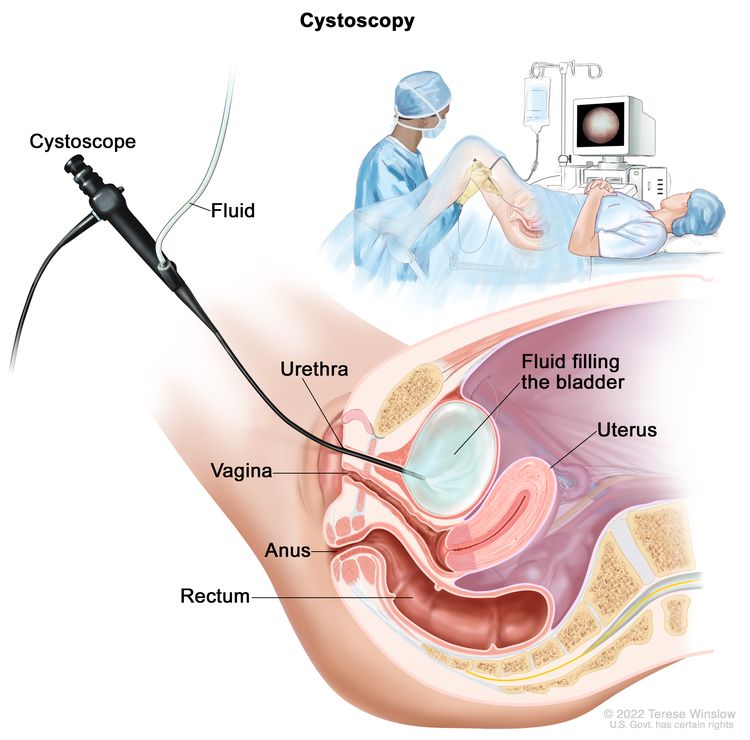
Cistoscopia. Se introduce un cistoscopio (instrumento delgado en forma de tubo, con una luz y una lente para observar) a través de la uretra hasta la vejiga. Se usa un líquido para llenar la vejiga. El médico observa una imagen de la pared interna de la vejiga en el monitor de una computadora para verificar si hay zonas anómalas.
- Proctoscopia: procedimiento para observar el interior del recto y del ano con un proctoscopio y determinar si hay áreas anormales. Un proctoscopio es un instrumento delgado en forma de tubo con una luz y una lente para observar el interior del recto y del ano. También puede tener una herramienta para extraer muestras de tejido, que se examinan bajo un microscopio para detectar signos de cáncer.
- La biopsia: es posible que se haga una biopsia para determinar si el cáncer se ha diseminado al cuello uterino. Se extrae una muestra de tejido del cuello uterino y se observa al microscopio. Generalmente, cuando se extrae solo una pequeña cantidad de tejido, la biopsia se realiza en el consultorio del médico. La biopsia de cono (extracción de un trozo de tejido más grande y en forma de cono del cuello uterino y del canal cervical) a menudo se lleva a cabo en el hospital. También se puede realizar una biopsia de la vulva para ver si el cáncer se ha propagado allí.
There are three ways that cancer spreads in the body.
Cancer can spread through tissue, the lymph system, and the blood:
- Tissue. The cancer spreads from where it began by growing into nearby areas.
- Lymph system. The cancer spreads from where it began by getting into the lymph system. The cancer travels through the lymph vessels to other parts of the body.
- Blood. The cancer spreads from where it began by getting into the blood. The cancer travels through the blood vessels to other parts of the body.
El cáncer puede extenderse desde donde comenzó a otras partes del cuerpo.
Cuando el cáncer se extiende a otra parte del cuerpo se denomina metástasis. Las células cancerosas se desprenden de donde comenzaron (tumor primario) y viajan a través del sistema linfático o la sangre.
- Sistema linfático: el cáncer entra en el sistema linfático, viaja a través de los vasos linfáticos y forma un tumor (tumor metastásico) en otra parte del cuerpo.
- Sangre: el cáncer llega a la sangre, viaja a través de los vasos sanguíneos y forma un tumor (tumor metastásico) en otra parte del cuerpo.
El tumor metastásico es del mismo tipo de cáncer que el tumor primario. Por ejemplo, si el cáncer de vagina se propaga al pulmón, las células cancerosas en el pulmón son en realidad células cancerosas de vagina. La enfermedad es cáncer vaginal metastásico, no cáncer de pulmón.
En la neoplasia intraepitelial vaginal (NIV) se encuentran células anómalas en el tejido que reviste el interior de la vagina.
Estas células anómalas no son cancerosas. La neoplasia intraepitelial vaginal (NIV) se clasifica según la profundidad a la que se encuentran las células anómalas en el tejido que recubre la vagina:
- NIV 1: las células anómalas se encuentran en el tercio más externo del tejido que recubre la vagina.
- NIV 2: las células anómalas se encuentran en los dos tercios más externos del tejido que recubre la vagina.
- NIV 3: las células anómalas se encuentran en más de dos tercios del tejido que recubre la vagina. Cuando las lesiones de NIV 3 se encuentran en todo el espesor del tejido que recubre la vagina, se denomina carcinoma in situ.
La neoplasia intraepitelial vaginal puede convertirse en cáncer y extenderse a la pared vaginal.
Para clasificar el cáncer de vagina se utilizan los siguientes estadios:
Estadio I
En el estadio I, el cáncer se encuentra solo en la pared de la vagina.
Estadio II
En el estadio II, el cáncer se ha diseminado a través de la pared de la vagina hasta el tejido que la rodea. El cáncer no se ha diseminado a la pared de la pelvis.
Estadio III
En el estadio III, el cáncer se ha diseminado a la pared de la pelvis.
Estadio IV
El estadio IV se divide en estadio IVA y estadio IVB:
- Estadio IVA: Cáncer puede haberse propagado a una o más de las siguientes áreas:
- Estadio IVB: el cáncer se ha diseminado a partes del cuerpo que no están cerca de la vagina, como los pulmones o los huesos.
El cáncer de vagina puede recidivar (regresar) después de haber sido tratado.
El cáncer puede reaparecer en la vagina o en otras partes del cuerpo.
Treatment Option Overview
Puntos clave
- Existen diferentes tipos de tratamiento para las pacientes con cáncer de vagina.
- The following types of treatment are used:
- Cirugía
- Radioterapia
- Quimioterapia
- New types of treatment are being tested in clinical trials.
- Inmunoterapia
- Radiosensibilizadores
- El tratamiento para el cáncer de vagina puede causar efectos secundarios.
- Patients may want to think about taking part in a clinical trial.
- Patients can enter clinical trials before, during, or after starting their cancer treatment.
- Pueden ser necesarias pruebas de seguimiento.
Existen diferentes tipos de tratamiento para las pacientes con cáncer de vagina.
Existen diferentes tipos de tratamientos para pacientes con cáncer vaginal . Algunos son tratamientos estándar (los que se utilizan actualmente) y otros se están probando en ensayos clínicos . Un ensayo clínico es un estudio de investigación cuyo objetivo es mejorar los tratamientos actuales u obtener información sobre nuevos tratamientos para pacientes con cáncer . Cuando los ensayos clínicos demuestran que un nuevo tratamiento es mejor que el estándar, este puede convertirse en el tratamiento estándar. Las pacientes pueden considerar participar en un ensayo clínico. Algunos ensayos clínicos están abiertos únicamente a pacientes que aún no han comenzado el tratamiento.
The following types of treatment are used:
Cirugía
La cirugía es una opción de tratamiento tanto para la neoplasia intraepitelial vaginal (VaIN) como para el cáncer vaginal.
Los siguientes tipos de cirugía pueden usarse para tratar la NIV:
- Cirugía láser: procedimiento quirúrgico que utiliza un rayo láser (haz angosto de luz intensa) como si fuera un bisturí para realizar cortes sin sangre en el tejido o para extirpar una lesión superficial, por ejemplo, un tumor.
- Escisión local amplia: procedimiento quirúrgico que extirpa el cáncer y parte del tejido sano que lo rodea.
- Vaginectomía: cirugía para extirpar la totalidad o una parte de la vagina. Es posible que se necesiten injertos de piel de otras partes del cuerpo para reconstruir la vagina.
Para el tratamiento del cáncer de vagina se utilizan los siguientes tipos de cirugía:
- Escisión local amplia: procedimiento quirúrgico que extirpa el cáncer y parte del tejido sano que lo rodea.
- Vaginectomía: cirugía para extirpar la totalidad o una parte de la vagina. Es posible que se necesiten injertos de piel de otras partes del cuerpo para reconstruir la vagina.
- Histerectomía total: cirugía para extirpar el útero, incluido el cuello uterinoCuando el útero y el cuello uterino se extirpan a través de la vagina, la cirugía se llama vaginal histerectomía. Si el útero y el cuello uterino se extraen a través de una gran incisión (corte) en el abdomen, la operación se llama total abdominal histerectomía. Si se extraen el útero y el cuello uterino a través de una pequeña incisión en el abdomen usando un laparoscopio, la operación se llama total laparoscópica histerectomía.
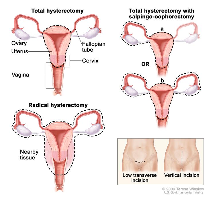
Histerectomía. El útero se extirpa quirúrgicamente con o sin otros órganos o tejidos. En una histerectomía total, se extirpan el útero y el cuello uterino. En una histerectomía total con salpingooforectomía, (a) se extirpan el útero, un ovario y una trompa de Falopio (unilaterales) o (b) se extirpan el útero, ambos ovarios y trompas de Falopio (bilaterales). En una histerectomía radical, se extirpan el útero, el cuello uterino, ambos ovarios, ambas trompas de Falopio y el tejido circundante. Estos procedimientos se realizan mediante una incisión transversal baja o una incisión vertical.
- Disección de ganglios linfáticos: Procedimiento quirúrgico en el que se extirpan los ganglios linfáticos y se examina una muestra de tejido al microscopio para detectar signos de cáncer. Este procedimiento también se denomina linfadenectomía. Si el cáncer se localiza en la parte superior de la vagina, se pueden extirpar los ganglios linfáticos pélvicos . Si se localiza en la parte inferior de la vagina, se pueden extirpar los ganglios linfáticos de la ingle .
- Exenteración pélvica: cirugía para extirpar la parte inferior del colon, el recto, la vejiga, el cuello uterino, la vagina y los ovarios. También se extirpan los ganglios linfáticos cercanos. Se hacen aberturas artificiales (estoma) para que la orina y la materia fecal pasen del cuerpo a una bolsa recolectora.
Después de que el médico extirpe todo el cáncer visible en el momento de la cirugía, algunos pacientes pueden recibir radioterapia para eliminar cualquier célula cancerosa que haya podido quedar. El tratamiento que se administra después de la cirugía para reducir el riesgo de que el cáncer reaparezca se denomina terapia adyuvante.
Radioterapia
La radioterapia es un tratamiento contra el cáncer que utiliza rayos X de alta energía u otros tipos de radiación para matar las células cancerosas o evitar que crezcan. Hay dos tipos de radioterapia:
- La radioterapia externa utiliza una máquina que envía radiación desde el exterior del cuerpo hacia la zona donde se encuentra el cáncer.
- La radioterapia interna utiliza una sustancia radiactiva sellada en agujas, semillas, cables o catéteres que se colocan directamente en el cáncer o cerca de él.
La forma en que se administra la radioterapia depende del tipo y estadio del cáncer que se está tratando. Tanto la radioterapia externa como la interna se usan para tratar el cáncer de vagina y, a veces, también como terapia paliativa para aliviar los síntomas y mejorar la calidad de vida.
Quimioterapia
La quimioterapia es un tratamiento contra el cáncer que utiliza fármacos para detener el crecimiento de las células cancerosas, ya sea destruyéndolas o impidiendo su división. Cuando la quimioterapia se administra por vía oral o mediante inyección intravenosa o intramuscular, los fármacos entran en el torrente sanguíneo y pueden afectar a las células cancerosas en todo el cuerpo ( quimioterapia sistémica ). Cuando la quimioterapia se administra directamente en el líquido cefalorraquídeo, un órgano o una cavidad corporal como el abdomen, los fármacos afectan principalmente a las células cancerosas de esas zonas ( quimioterapia regional ). La forma de administración de la quimioterapia depende del tipo y estadio del cáncer que se esté tratando.
Para el cáncer vaginal escamocelular, se puede aplicar quimioterapia tópica en la vagina en forma de crema o loción.
New types of treatment are being tested in clinical trials.
En esta sección se resumen los tratamientos que se están estudiando en ensayos clínicos. Es posible que no se mencionen todos los tratamientos nuevos que se están estudiando. La información sobre los ensayos clínicos está disponible en el sitio web del NCI.
Inmunoterapia
La inmunoterapia es un tratamiento que utiliza el sistema inmunológico del paciente para combatir el cáncer. Las sustancias producidas por el cuerpo o en un laboratorio se utilizan para estimular, dirigir o restaurar las defensas naturales del cuerpo contra el cáncer.
El imiquimod es un modificador de la respuesta inmune que se está estudiando para tratar lesiones vaginales. Se aplica sobre la piel en forma de crema.
Radiosensibilizadores
Los radiosensibilizadores son medicamentos que aumentan la sensibilidad de las células tumorales a la radioterapia. Combinar radioterapia con radiosensibilizadores puede destruir más células tumorales.
El tratamiento para el cáncer de vagina puede causar efectos secundarios.
For information about side effects caused by treatment for cancer, visit our Side Effects page.
Patients may want to think about taking part in a clinical trial.
For some patients, taking part in a clinical trial may be the best treatment choice. Clinical trials are part of the cancer research process. Clinical trials are done to find out if new cancer treatments are safe and effective or better than the standard treatment.
Many of today's standard treatments for cancer are based on earlier clinical trials. Patients who take part in a clinical trial may receive the standard treatment or be among the first to receive a new treatment.
Patients who take part in clinical trials also help improve the way cancer will be treated in the future. Even when clinical trials do not lead to effective new treatments, they often answer important questions and help move research forward.
Patients can enter clinical trials before, during, or after starting their cancer treatment.
Some clinical trials only include patients who have not yet received treatment. Other trials test treatments for patients whose cancer has not gotten better. There are also clinical trials that test new ways to stop cancer from recurring (coming back) or reduce the side effects of cancer treatment.
Clinical trials are taking place in many parts of the country. Information about clinical trials supported by NCI can be found on NCI’s clinical trials search webpage. Clinical trials supported by other organizations can be found on the ClinicalTrials.gov website.
Pueden ser necesarias pruebas de seguimiento.
Es posible que se repitan algunas de las pruebas que se realizaron para diagnosticar o para determinar el estadio del cáncer. Algunas pruebas se repetirán para ver si el tratamiento está funcionando. Los resultados de estas pruebas pueden usarse para decidir si continuar, cambiar o suspender el tratamiento.
Tratamiento de la neoplasia intraepitelial vaginal (NIV)
For information about the treatments listed below, see the Treatment Option Overview section.
El tratamiento de la neoplasia intraepitelial vaginal (NIV) puede incluir las siguientes opciones:
- Cirugía ( cirugía láser después de una biopsia ).
- Cirugía (escisión local amplia) con injerto de piel
- Cirugía (vaginectomía parcial o total) con o sin injerto de piel
- Quimioterapia tópica.
- Radioterapia interna.
- Ensayo clínico de inmunoterapia ( imiquimod ) aplicada sobre la piel.
Tratamiento del cáncer de vagina en estadio I
For information about the treatments listed below, see the Treatment Option Overview section.
El tratamiento de las lesiones causadas por cáncer vaginal escamocelular en estadio I que tienen menos de 0.5 centímetros de grosor puede incluir las siguientes alternativas:
- Radioterapia externa, especialmente para tumores grandes o ganglios linfáticos cercanos a tumores en la parte inferior de la vagina
- Radioterapia interna.
- Cirugía ( escisión local amplia o vaginectomía con reconstrucción vaginal ). Se puede administrar radioterapia después de la cirugía.
El tratamiento de las lesiones causadas por cáncer vaginal escamocelular en estadio I que tienen más de 0.5 centímetros de grosor puede incluir las siguientes alternativas:
- Cirugía:
- Vaginectomía y disección de ganglios linfáticos con o sin reconstrucción vaginal para las lesiones en el tercio superior de la vagina
- Disección de ganglios linfáticos para las lesiones en el tercio inferior de la vagina
- Después de la cirugía, es posible que se administre alguna de las siguientes opciones de radioterapia:
- Radioterapia externa con o sin radioterapia interna
- Radioterapia interna
- Para las lesiones en el tercio inferior de la vagina, se puede administrar radioterapia en los ganglios linfáticos cercanos al tumor.
El tratamiento del adenocarcinoma vaginal en estadio I puede incluir las siguientes opciones:
- Cirugía ( vaginectomía e histerectomía con disección de ganglios linfáticos ). Posteriormente, puede realizarse una reconstrucción vaginal y/o radioterapia .
- Radioterapia interna. También se puede administrar radioterapia externa a los ganglios linfáticos cercanos a los tumores en la parte inferior de la vagina.
- Una combinación de terapias que puede incluir la escisión local amplia con o sin disección de ganglios linfáticos y la radioterapia interna.
Tratamiento del cáncer de vagina en estadio II, estadio III y estadio IVA
For information about the treatments listed below, see the Treatment Option Overview section.
El tratamiento del cáncer de vagina en estadio II, estadio III y estadio IVA es el mismo para el carcinoma escamocelular y el adenocarcinoma. El tratamiento puede incluir las siguientes alternativas:
- Radioterapia interna y/o externa en la vagina . También se puede administrar radioterapia a los ganglios linfáticos cercanos a los tumores en la parte inferior de la vagina .
- Cirugía ( vaginectomía o exenteración pélvica ) con o sin radioterapia.
- Quimioterapia administrada junto con radioterapia.
Tratamiento del cáncer de vagina en estadio IVB
For information about the treatments listed below, see the Treatment Option Overview section.
El tratamiento del cáncer de vagina en estadio IVB es el mismo para el carcinoma escamocelular y el adenocarcinoma. El tratamiento puede incluir las siguientes opciones:
- La radioterapia se utiliza como terapia paliativa para aliviar los síntomas y mejorar la calidad de vida . También se puede administrar quimioterapia .
Aunque no se ha demostrado que ningún fármaco anticancerígeno prolongue la vida de las pacientes con cáncer vaginal en estadio IVB, a menudo se las trata con regímenes utilizados para el cáncer de cuello uterino . Para más información, consulte Tratamiento del cáncer de cuello uterino .
Tratamiento del cáncer de vagina recidivante
For information about the treatments listed below, see the Treatment Option Overview section.
El tratamiento del cáncer de vagina recidivante puede incluir los siguientes opciones:
Aunque no se ha demostrado que ningún fármaco anticancerígeno prolongue la vida de las pacientes con cáncer vaginal recurrente, a menudo se las trata con regímenes utilizados para el cáncer de cuello uterino . Para más información, consulte Tratamiento del cáncer de cuello uterino .
Puede utilizar la búsqueda de ensayos clínicos y encontrar ensayos clínicos sobre cáncer patrocinados por el NCI que acepten participantes. La búsqueda le permite filtrar los ensayos según el tipo de cáncer, la edad y el lugar donde se realizan los ensayos. También encontrará información general sobre los ensayos clínicos.
Más información sobre el cáncer de vagina
Para obtener más información del National Cancer Institute sobre el cáncer vaginal, consulte los siguientes sitios web:
- Página de inicio sobre el cáncer de vagina
- ¿Qué es el cáncer de cuello uterino?
- Terapia con láser para tratar el cáncer
- Tomografía computarizada para el cáncer
- Inmunomoduladores
- El virus del papiloma humano (VPH) y el cáncer
For general cancer information and other resources from the National Cancer Institute, visit:
Sobre este resumen del PDQ
Acerca del PDQ
El Physician Data Query (PDQ) es la base de datos integral sobre el cáncer del National Cancer Institute (NCI). La base de datos del PDQ contiene resúmenes con la última información publicada sobre prevención, detección, genética, tratamiento, atención médica de apoyo y medicina complementaria y alternativa relacionada con el cáncer. La mayoría de los resúmenes se presentan en dos versiones. Las versiones para profesionales de la salud contienen información detallada escrita en lenguaje técnico. Las versiones para pacientes están escritas en un lenguaje fácil de entender y no tan técnico. Ambas versiones contienen información precisa y actualizada sobre el cáncer. La mayoría de las versiones también están disponibles en español.
El PDQ es un servicio del NCI. El NCI es parte de los Institutos Nacionales de Salud (NIH), que son el centro de investigación biomédica del Gobierno federal. Los resúmenes del PDQ se basan en una revisión independiente de la literatura médica. No son declaraciones de políticas del NCI ni de los NIH.
Propósito de este resumen
Este resumen del PDQ sobre el cáncer contiene información actualizada del tratamiento del cáncer de vagina. El propósito es informar y ayudar a los pacientes, sus familiares y cuidadores. No da pautas ni recomendaciones formales para tomar decisiones relacionadas con la atención médica.
Revisores y actualizaciones
Los comités editoriales escriben los resúmenes de información sobre el cáncer del PDQ y los mantienen actualizados. Estos comités están formados por equipos de especialistas en el tratamiento del cáncer y otras especialidades relacionadas con esta enfermedad. Los resúmenes se revisan periódicamente y se modifican cuando hay información nueva. La fecha de actualización al pie de cada resumen indica cuándo se realizó el cambio más reciente.
The information in this patient summary was taken from the health professional version, which is reviewed regularly and updated as needed, by the PDQ Adult Treatment Editorial Board.
Información sobre ensayos clínicos
Un ensayo clínico es un estudio para responder a una pregunta científica como, por ejemplo, si un tratamiento es mejor que otro. Los ensayos se basan en estudios anteriores y en lo aprendido en el laboratorio. Cada ensayo responde a determinadas preguntas científicas que permiten encontrar nuevas y mejores formas de ayudar a los pacientes con cáncer. Durante los ensayos clínicos de tratamiento, se recopila información sobre los efectos de un nuevo tratamiento y su eficacia. Si un ensayo clínico demuestra que un nuevo tratamiento es mejor que uno que se utiliza actualmente, el nuevo tratamiento puede convertirse en “estándar”. Los pacientes pueden valorar la posibilidad de participar en un ensayo clínico. Algunos ensayos clínicos solo están abiertos a pacientes que no hayan iniciado el tratamiento.
Los ensayos clínicos se pueden encontrar en línea en el sitio web del NCI. Para obtener más información, llame al Servicio de Información sobre el Cáncer (CIS, por sus siglas en inglés), el centro de contacto del NCI, al 1-800-4-CANCER (1-800-422-6237).
Permiso de uso de este resumen
Physician Data Query (PDQ) es una marca registrada. Se autoriza el libre uso del contenido de los documentos del PDQ como texto. Sin embargo, no se podrá identificar como un resumen de información sobre cáncer del PDQ del NCI, salvo que se reproduzca en su totalidad y se actualice con regularidad. Por otra parte, se permite que los autores incluyan una oración como “en el resumen del PDQ del NCI sobre la prevención del cáncer de mama se describen, de manera concisa, los siguientes riesgos: [incluir fragmento del resumen]”.
La forma recomendada para citar este resumen del PDQ es:
Comité editorial del PDQ® sobre el tratamiento para adultos. Tratamiento del cáncer de vagina (PDQ). Bethesda, MD: National Cancer Institute. Actualizado el [DD/MM/AAAA].
Las imágenes de este resumen se utilizan con el permiso del autor, artista y/o editorial para uso exclusivo en los resúmenes del PDQ. Si desea usar una imagen de un resumen del PDQ sin incluir el resumen completo, debe obtener autorización del propietario. El National Cancer Institute no puede otorgar dicho permiso. Para obtener más información sobre el uso de las imágenes de este resumen o de otras ilustraciones relacionadas con el cáncer, consulte Visuals Online, una colección de más de 3,000 imágenes científicas.
Descargo de responsabilidad
La información de estos resúmenes no debe utilizarse para tomar decisiones sobre reembolsos de seguros. Puede encontrar más información sobre la cobertura de seguros en Cancer.gov en el sitio Manejo de la atención del cáncer.
Contáctenos
Puede encontrar más información sobre cómo contactarnos o recibir ayuda en el sitio web Cancer.gov en la página Comuníquese con el NCI. También puede enviar sus preguntas a Cancer.gov en el apartado Escríbanos del sitio web.
Actualizado:
URL de origen: https://www.cancer.gov/node/5268/syndication
Agencia de origen: National Cancer Institute (NCI)
Fecha de captura: 14/09/2013 09:02:44.0