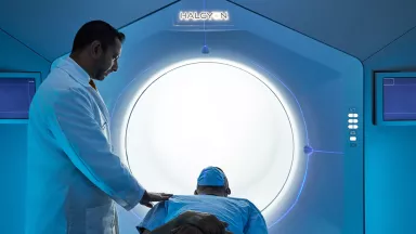
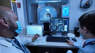
Acceda a una atención excepcional para el sarcoma de tejidos blandos en Montefiore Einstein Comprehensive Cancer Center. Nuestro centro del cáncer fue uno de los primeros en recibir la designación del NCI en el país y, por más de 50 años, hemos sido líderes en la investigación, el diagnóstico y el tratamiento de más de 200 tipos de cáncer. Nuestros especialistas en cáncer de renombre mundial atienden a más de 5500 nuevos pacientes cada año. Nuestros excepcionales equipos ofrecen tratamiento médico y quirúrgico para el sarcoma de tejidos blandos, y combinan su experiencia para brindar una atención integral en un único entorno clínico.
Recibirá una atención compasiva y personalizada que cumple con los más altos estándares de calidad y seguridad. El tratamiento contra el sarcoma de tejidos blandos del Montefiore Einstein Comprehensive Cancer Center es uno de los más completos del país, y ofrece tratamiento oncológico médico y quirúrgico, así como terapia dirigida.
Cada semana, nuestro equipo colabora con especialistas en radiología, patología, oncología médica, oncología radioterápica y oncología quirúrgica, lo que permite el diseño de tratamientos personalizados específicos para cada tipo de cáncer.
Cuando necesite atención para el sarcoma de tejidos blandos, recurra a nuestros proveedores, quienes se dedican con pasión a combatir el cáncer y a atender todas sus necesidades de salud.
El Montefiore Einstein Comprehensive Cancer Center, designado como centro integral del cáncer por el National Cancer Institute (NCI), apoya la misión y las normas del NCI. La siguiente información sobre los tipos de cáncer, prevención y tratamientos ha sido facilitada por el NCI.
Tratamiento del sarcoma de tejidos blandos (PDQ®): versión para pacientes
Información general sobre el sarcoma de tejidos blandos
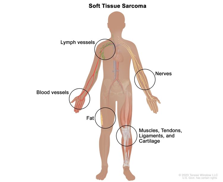
El sarcoma de tejidos blandos se forma en los tejidos blandos del cuerpo, incluidos los músculos, tendones, ligamentos, cartílagos, grasa, vasos sanguíneos, vasos linfáticos, nervios y tejidos alrededor de las articulaciones.
Existen muchos tipos de sarcoma de tejidos blandos. Las células de cada tipo de sarcoma se ven diferentes al microscopio, según el tipo de tejido blando en el que se haya originado el cáncer.
Para obtener más información sobre los sarcomas de tejidos blandos, consulte los siguientes enlaces:
Tener ciertos trastornos hereditarios puede aumentar el riesgo de tener sarcoma de tejidos blandos.
Todo lo que aumenta la probabilidad de que una persona padezca una enfermedad se denomina factor de riesgo. No todas las personas con uno o más de estos factores de riesgo desarrollarán sarcoma de tejidos blandos, y este se presentará en personas sin factores de riesgo conocidos. Consulte con su médico si cree que puede estar en riesgo. Los factores de riesgo del sarcoma de tejidos blandos incluyen los siguientes trastornoshereditarios:
Uno de los signos del sarcoma de tejido blando es un bulto o hinchazón en el tejido blando del cuerpo.
Un sarcoma puede aparecer como un bulto indoloro debajo de la piel, a menudo en un brazo o una pierna. Los sarcomas que se originan en el abdomen pueden no presentar signos ni síntomas hasta que alcanzan un gran tamaño. A medida que el sarcoma crece y presiona órganos, nervios, músculos o vasos sanguíneos cercanos, se presentan los siguientes signos y síntomas:
Dolor
Dificultad para respirar
Otras afecciones pueden causar los mismos signos y síntomas. Consulte con su médico si presenta alguno de estos problemas.
El sarcoma de tejidos blandos se diagnostica mediante una biopsia.
Si su médico sospecha que puede tener un sarcoma de tejidos blandos, le realizará una biopsia. El tipo de biopsia dependerá del tamaño del tumor y su ubicación en el cuerpo. Se pueden realizar los siguientes tipos de biopsia:
Biopsia incisional: extracción de parte de un bulto o de una muestra de tejido. Se puede realizar una biopsia incisional cuando no es seguro realizar una biopsia con aguja gruesa o los resultados de la biopsia con aguja gruesa no son claros.
La planificación cuidadosa de la biopsia debe involucrar al cirujano, un oncólogo radioterapeuta y un radiólogo intervencionista que utiliza imágenes médicas para guiar el diagnóstico. Se tomarán muestras del tumor primario, los ganglios linfáticos y otras áreas sospechosas. Un patólogo observará el tejido bajo un microscopio para buscar células cancerosas y determinar el grado del tumor. El grado de un tumor depende de qué tan anormales se vean las células cancerosas bajo un microscopio y con qué rapidez se multiplican. Los tumores de grado alto generalmente crecen y se propagan más rápidamente que los tumores de bajo grado.
Debido a que el sarcoma de tejido blando puede ser difícil de diagnosticar, las muestras de tejido deben ser examinadas por un patólogo que tenga experiencia en el diagnóstico de sarcoma de tejido blando.
Se pueden realizar las siguientes pruebas en el tejido extraído:
Inmunohistoquímica: prueba de laboratorio que utiliza anticuerpos para detectar ciertos antígenos (marcadores) en una muestra de tejido de un paciente. Los anticuerpos suelen estar unidos a una enzima o un tinte fluorescente. Después de que los anticuerpos se unen a un antígeno específico en la muestra de tejido, la enzima o el tinte se activa y el antígeno se puede observar bajo un microscopio. Este tipo de prueba se usa para diagnosticar el cáncer y diferenciarlo de otros tipos de cáncer.
Citometría de flujo: prueba de laboratorio que mide la cantidad de células en una muestra, el porcentaje de células vivas en una muestra y ciertas características de las células, como el tamaño, la forma y la presencia de marcadores tumorales (u otros) en la superficie celular. Las células de una muestra de sangre, médula ósea u otro tejido de un paciente se tiñen con un tinte fluorescente, se colocan en un líquido y luego se pasan una a una a través de un haz de luz. Los resultados de la prueba se basan en cómo reaccionan las células teñidas con el tinte fluorescente al haz de luz.
Perfil molecular: método de laboratorio que utiliza una muestra de tejido, sangre u otro líquido corporal para detectar determinados genes, proteínas u otras moléculas que pueden ser un signo de una enfermedad o afección, como el cáncer. También se puede utilizar para detectar determinados cambios en un gen o cromosoma que pueden aumentar el riesgo de una persona de desarrollar cáncer u otras enfermedades. Se puede realizar junto con otros procedimientos, como biopsias, para diagnosticar algunos tipos de cáncer. También se puede utilizar para planificar el tratamiento, averiguar qué tan bien está funcionando el tratamiento, hacer un pronóstico o predecir si el cáncer regresará o se propagará a otras partes del cuerpo.
Microscopíaóptica y electrónica: prueba de laboratorio en la que se observan las células de una muestra de tejido bajo microscopios regulares y de alta potencia para buscar ciertos cambios en las células.
Análisis citogenético: prueba de laboratorio en la que se cuentan los cromosomas de las células de una muestra de tejido y se comprueba si hay cambios, como cromosomas rotos, faltantes, reorganizados o adicionales. Los cambios en ciertos cromosomas pueden ser un signo de cáncer. El análisis citogenético se utiliza para diagnosticar el cáncer, planificar el tratamiento o determinar qué tan bien está funcionando el tratamiento.
Hibridación in situ con fluorescencia (FISH): prueba de laboratorio que se utiliza para observar y contar genes o cromosomas en células y tejidos. Se generan fragmentos de ADN con colorantes fluorescentes en el laboratorio y se añaden a una muestra de células o tejidos del paciente. Cuando estos fragmentos de ADN teñidos se unen a ciertos genes o zonas de cromosomas en la muestra, se iluminan al observarlos con un microscopio de fluorescencia. La prueba FISH se utiliza para diagnosticar el cáncer y planificar el tratamiento.
Ciertos factores afectan las opciones de tratamiento y el pronóstico (es decir, la posibilidad de recuperación).
Las opciones de tratamiento y el pronóstico dependen de los siguientes aspectos:
Los tumores pequeños y de bajo grado, especialmente en el tronco, los brazos o las piernas, frecuentemente se tratan solo con cirugía. Los sarcomas de alto grado son más difíciles de tratar y tienen más probabilidades de propagarse.
Estadios del sarcoma de tejido blando
Puntos clave
Una vez que se diagnostica el sarcoma de tejidos blandos, se realizan pruebas para determinar si las células cancerosas se han extendido dentro del tejido blando o hacia otras partes del cuerpo.
El cáncer se propaga por el cuerpo de tres maneras.
El cáncer puede extenderse desde donde comenzó a otras partes del cuerpo.
El grado del tumor también se utiliza para describir el cáncer y planificar el tratamiento.
Para clasificar el sarcoma de tejidos blandos del tronco, los brazos y las piernas, se utilizan los siguientes estadios:
Estadio I
Estadio II
Estadio III
Estadio IV
Para clasificar el sarcoma de tejido blando del retroperitoneo, se usan los siguientes estadios:
Estadio I
Estadio II
Estadio III
Estadio IV
No existe un sistema de estadificación estándar para el sarcoma de tejidos blandos de la cabeza, el cuello, el tórax y el abdomen.
El sarcoma de tejidos blandos puede recidivar (regresar) después de haber sido tratado.
Una vez que se diagnostica el sarcoma de tejidos blandos, se realizan pruebas para determinar si las células cancerosas se han extendido dentro del tejido blando o hacia otras partes del cuerpo.
El proceso que se utiliza para determinar si el cáncer se ha propagado dentro del tejido blando o a otras partes del cuerpo se denomina estadificación. La estadificación del sarcoma de tejido blando también se basa en el grado y el tamaño del tumor, y en si se ha propagado a los ganglios linfáticos o a otras partes del cuerpo. La información obtenida del proceso de estadificación determina el estadio de la enfermedad. Es importante conocer el estadio para planificar el tratamiento.
Además de preguntarle sobre su historial de salud familiar y personal, y de hacerle un examen físico, su médico puede realizar las siguientes pruebas y procedimientos durante el proceso de estadificación:
Radiografía del tórax: radiografía de los órganos y huesos del interior del tórax. Los rayos X son un tipo de haz de energía que puede atravesar el cuerpo, plasmarse en una película y generar una imagen de áreas del interior del cuerpo.
Estudios de química sanguínea: procedimiento mediante el cual se analiza una muestra de sangre para medir la cantidad de ciertas sustancias que los órganos y tejidos del cuerpo liberan en la sangre. Una cantidad inusual (superior o inferior a la normal) de una sustancia puede ser un signo de enfermedad.
Hemograma completo: procedimiento en el que se extrae una muestra de sangre y se examina:
La parte de la muestra de sangre compuesta por glóbulos rojos.
Tomografía computarizada (TC): procedimiento que genera una serie de imágenes detalladas de áreas internas del cuerpo, como los pulmones, el abdomen y la pelvis, tomadas desde diferentes ángulos. Las imágenes son tomadas por una computadora conectada a una máquina de rayos X. Se puede inyectar un tinte en una vena o administrarlo por vía oral para que los órganos o tejidos se vean más claramente. Este procedimiento también se llama tomografía axial computarizada.
Resonancia magnética (RM): se utilizan un imán, ondas de radio y una computadora para generar imágenes detalladas de las estructuras internas del cuerpo. Este procedimiento también se denomina resonancia magnética nuclear (RMN).
Tomografía por emisión de positrones (PET): procedimiento para detectar células tumorales malignas en el cuerpo. Se inyecta una pequeña cantidad de glucosaradiactiva (azúcar) en una vena. El escáner PET gira alrededor del cuerpo y genera una imagen de las zonas donde se utiliza la glucosa. Las células tumorales malignas se ven más brillantes en la imagen porque son más activas y absorben más glucosa que las células normales.
Los resultados de estas pruebas se estudian junto con los resultados de la biopsia del tumor para determinar el estadio del sarcoma de tejido blando antes de administrar el tratamiento. A veces se administra quimioterapia o radioterapia como tratamiento inicial y luego se vuelve a estadificar el sarcoma de tejido blando.
El cáncer se propaga por el cuerpo de tres maneras.
Tejidos: el cáncer se extiende desde el lugar donde comenzó y crece hacia las zonas circundantes.
Sistema linfático: el cáncer se extiende desde el lugar donde comenzó hacia el sistema linfático. El cáncer viaja a través de los vasos linfáticos a otras partes del cuerpo.
Sangre: el cáncer se extiende desde el lugar donde comenzó hacia la sangre. El cáncer viaja a través de los vasos sanguíneos a otras partes del cuerpo.
El cáncer puede extenderse desde donde comenzó a otras partes del cuerpo.
Cuando el cáncer se extiende a otra parte del cuerpo se denomina metástasis. Las células cancerosas se desprenden de donde comenzaron (tumor primario) y viajan a través del sistema linfático o la sangre.
Sistema linfático: el cáncer entra en el sistema linfático, viaja a través de los vasos linfáticos y forma un tumor (tumor metastásico) en otra parte del cuerpo.
Sangre: el cáncer llega a la sangre, viaja a través de los vasos sanguíneos y forma un tumor (tumor metastásico) en otra parte del cuerpo.
El tumor metastásico es el mismo tipo de cáncer que el tumor primario. Por ejemplo, si el sarcoma de tejido blando se extiende al pulmón, las células cancerosas en el pulmón son en realidad células de sarcoma de tejido blando. La enfermedad es sarcoma de tejido blando metastásico, no cáncer de pulmón.
Muchas muertes por cáncer se producen cuando el cáncer se desplaza del tumor original y se extiende a otros tejidos y órganos. Esto se llama cáncer metastásico. Esta animación muestra cómo las células cancerosas viajan desde el lugar donde se formaron por primera vez a otras partes del cuerpo.
El grado del tumor también se utiliza para describir el cáncer y planificar el tratamiento.
El grado del tumor describe el aspecto anormal de las células cancerosas bajo un microscopio y la rapidez con la que es probable que el tumor crezca y se extienda. El sarcoma de tejido blando se describe como de grado bajo, grado medio y grado alto:
Grado bajo: en el sarcoma de tejido blando de grado bajo, las células cancerosas se parecen más a las células normales bajo un microscopio, y crecen y se diseminan más lentamente que en el sarcoma de tejido blando de grados medio y alto.
Grado medio: en el sarcoma de tejido blando de grado medio, las células cancerosas se ven más anormales al microscopio, y crecen y se diseminan más rápidamente que en el sarcoma de tejido blando de grado bajo.
Grado alto: en el sarcoma de tejido blando de grado alto, las células cancerosas se ven más anormales bajo el microscopio, y crecen y se diseminan más rápidamente que en el sarcoma de tejido blando de grados bajo y medio.
Para clasificar el sarcoma de tejidos blandos del tronco, los brazos y las piernas, se utilizan los siguientes estadios:
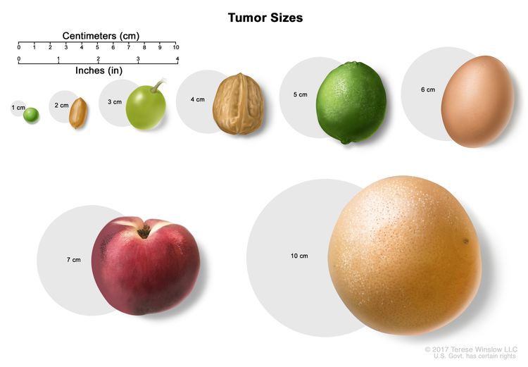
El tamaño de los tumores a menudo se mide en centímetros (cm) o pulgadas (in). A veces se usan alimentos comunes para mostrar el tamaño de un tumor en centímetros: una arveja o guisante (1 cm), un maní o cacahuate (2 cm), una uva (3 cm), una nuez (4 cm), una lima o limón verde (5 cm o 2 in), un huevo (6 cm), un durazno (7 cm) y un pomelo (10 cm o 4 in).
El tumor es de cualquier tamaño y de cualquier grado, y puede haberse extendido a los ganglios linfáticos cercanos. El cáncer se ha extendido a otras partes del cuerpo, como los pulmones.
Para clasificar el sarcoma de tejido blando del retroperitoneo, se usan los siguientes estadios:
El tamaño de los tumores a menudo se mide en centímetros (cm) o pulgadas (in). A veces se usan alimentos comunes para mostrar el tamaño de un tumor en centímetros: una arveja o guisante (1 cm), un maní o cacahuate (2 cm), una uva (3 cm), una nuez (4 cm), una lima o limón verde (5 cm o 2 in), un huevo (6 cm), un durazno (7 cm) y un pomelo (10 cm o 4 in).
No existe un sistema de estadificación estándar para el sarcoma de tejidos blandos de la cabeza, el cuello, el tórax y el abdomen.
El sarcoma de tejidos blandos puede recidivar (regresar) después de haber sido tratado.
El cáncer puede reaparecer en el mismo tejido blando o en otras partes del cuerpo.
Descripción general de las opciones de tratamiento
Puntos clave
Existen diferentes tipos de tratamiento para los pacientes con sarcoma de tejidos blandos.
Se utilizan los siguientes tipos de tratamiento:
Cirugía
Radioterapia
Quimioterapia
Terapia dirigida
Inmunoterapia
El tratamiento para el sarcoma de tejidos blandos puede causar efectos secundarios.
Los pacientes pueden evaluar la posibilidad de participar en un ensayo clínico.
Los pacientes pueden participar en ensayos clínicos antes, durante o después de comenzar el tratamiento contra el cáncer.
Pueden ser necesarias pruebas de seguimiento.
Existen diferentes tipos de tratamiento para los pacientes con sarcoma de tejidos blandos.
Existen diferentes tipos de tratamientos disponibles para los pacientes con sarcoma de tejidos blandos. Algunos tratamientos son estándar (el tratamiento que se utiliza actualmente) y otros se están probando en ensayos clínicos. Un ensayo clínico de tratamiento es un estudio de investigación cuyo objetivo es ayudar a mejorar los tratamientos actuales u obtener información sobre nuevos tratamientos para pacientes con cáncer. Cuando los ensayos clínicos muestran que un nuevo tratamiento es mejor que el tratamiento estándar, el nuevo tratamiento puede convertirse en el tratamiento estándar. Es posible que los pacientes quieran pensar en participar en un ensayo clínico. Algunos ensayos clínicos están abiertos solo a pacientes que no han comenzado el tratamiento.
Se utilizan los siguientes tipos de tratamiento:
Cirugía
La cirugía es el tratamiento más común para el sarcoma de tejidos blandos. Puede ser el único tratamiento necesario para tumores pequeños y de bajo grado, especialmente en el tronco, los brazos o las piernas. Se pueden utilizar los siguientes procedimientos quirúrgicos:
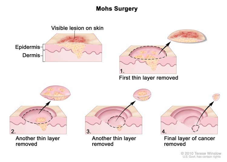
Cirugía micrográfica de Mohs: procedimiento en el que se corta el tumor de la piel en capas finas. Durante la cirugía, los bordes del tumor y cada capa del tumor extirpado se observan a través de un microscopio para comprobar si hay célulascancerosas. Se siguen eliminando capas hasta que no se ven más células cancerosas. Este tipo de cirugía conserva la mayor cantidad posible de tejidos sano y se usa a menudo donde la apariencia es importante, como en la piel. Ampliar
Escisión local amplia: extirpación del tumor junto con parte del tejido normal que lo rodea. Para los tumores de cabeza, cuello, abdomen y tronco, se extrae la menor cantidad de tejido normal posible.
Cirugía preservadora de una extremidad: extirpación del tumor en un brazo o pierna sin amputación, por lo que se salva el uso y apariencia de la extremidad. Es posible que primero se administre radioterapia o quimioterapia para reducir el tamaño del tumor. Luego se extirpa el tumor mediante una escisión local amplia. El tejido y el hueso que se extraen se pueden reemplazar con un injerto que utiliza tejido y hueso extraídos de otra parte del cuerpo del paciente o con un implante, como un hueso artificial.
Amputación: cirugía para extirpar parte o la totalidad de un brazo o una pierna. La amputación rara vez se usa para tratar el sarcoma de tejidos blandos.
Linfadenectomía: procedimiento quirúrgico en el que se extirpan los ganglios linfáticos y se examina una muestra de tejido bajo un microscopio para detectar signos de cáncer. Este procedimiento también se llama disección de ganglios linfáticos.
Se puede administrar radioterapia o quimioterapia antes o después de la cirugía para extirpar el tumor. Cuando se administran antes de la cirugía, la radioterapia o la quimioterapia reducirán el tamaño del tumor y la cantidad de tejido que se debe extirpar durante la cirugía. El tratamiento administrado antes de la cirugía se llama terapia neoadyuvante. Cuando se administran después de la cirugía para extirpar todo el tumor que se pueda ver, la radioterapia o la quimioterapia eliminarán las células cancerosas restantes. El tratamiento administrado después de la cirugía para reducir el riesgo de que el cáncer regrese se llama terapia adyuvante.
Radioterapia
La radioterapia es un tratamiento contra el cáncer que utiliza rayos X de alta energía u otros tipos de radiación para matar las células cancerosas o evitar que crezcan. Hay dos tipos de radioterapia:
Radioterapia externa se usa una máquina que envía radiación desde el exterior del cuerpo hacía el área en la que se encuentra el cáncer.
La radioterapia de intensidad modulada (RTIM) es un tipo de radioterapia tridimensional (3-D) que utiliza una computadora para generar imágenes del tamaño y la forma del tumor. Se dirigen rayos finos de radiación de diferentes intensidades (fuerzas) al tumor desde muchos ángulos. Este tipo de radioterapia externa causa menos daño al tejido sano circundante y es menos probable que cause sequedad en la boca, dificultad para tragar y daño a la piel.
La forma en que se administra la radioterapia depende del tipo y estadio del cáncer que se está tratando. Se puede usar radioterapia externa y radioterapia interna para tratar el sarcoma de tejido blando.
Quimioterapia
La quimioterapia es un tratamiento contra el cáncer que utiliza medicamentos para detener el crecimiento de las células cancerosas, ya sea destruyéndolas o impidiendo su división. Cuando la quimioterapia se administra por vía oral o se inyecta en una vena o músculo, los medicamentos entran en el torrente sanguíneo y pueden llegar a las células cancerosas de todo el cuerpo (quimioterapia sistémica).
La terapia dirigida es un tipo de tratamiento que utiliza medicamentos u otras sustancias para identificar y atacar células cancerosas específicas. Existen diferentes tipos de terapia dirigida, como los siguientes:
Terapia inhibidores de histona metiltransferasa: esta terapia puede ayudar a evitar que las células cancerosas crezcan. Tazemetostat es un tipo de terapia inhibidora de la histona metiltransferasa que se usa para tratar el sarcoma de tejido blando.
La inmunoterapia es un tratamiento en el que se utiliza el sistema inmunológico del paciente para combatir el cáncer. Las sustancias producidas por el cuerpo o en un laboratorio se utilizan para estimular, dirigir o restaurar las defensas naturales del cuerpo contra el cáncer.
La terapia con inhibidores de puntos de control inmunológico es un tipo de inmunoterapia. Algunos tipos de células del sistema inmunitario, como las células T, y algunas células cancerosas, tienen ciertas proteínas en su superficie, llamadas proteínas de punto de control, que mantienen bajo control las respuestas inmunitarias. Estos puntos de control ayudan a evitar que las respuestas inmunitarias sean demasiado fuertes y, en ocasiones, pueden impedir que las células T destruyan las células cancerosas. Cuando estos puntos de control están bloqueados, las células T pueden destruir mejor las células cancerosas.
Los tipos de terapia con inhibidores de puestos de control inmunitario son:
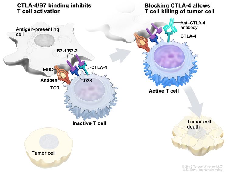
CTLA-4 terapia con inhibidores: CTLA-4 es una proteína en la superficie de las células T que ayuda a mantener bajo control las respuestas inmunitarias del cuerpo. Cuando la CTLA-4 se une a otra proteína llamada B7 en una célula cancerosa, impide que la célula T destruya la célula cancerosa. Los inhibidores de CTLA-4 se adhieren a la CTLA-4 y permiten que las células T eliminen las células cancerosas.
Ipilimumab es un tipo de inhibidor de la proteína CTLA-4 que se está estudiando para tratar el sarcoma de tejido blando.
Inhibidor de puestos de control inmunitario. Las proteínas de puestos de control, como la B7-1/B7-2 en las células presentadoras de antígenos (CPA) y CTLA-4 en los linfocitos T, ayudan a mantener bajo control la respuesta inmunitaria del cuerpo. Cuando el receptor de linfocitos T (TCR) se une al antígeno y a las proteínas del complejo mayor de histocompatibilidad (MHC) en las CPA, y la CD28 se une a la B7-1/B7-2 en las CPA, el linfocito T puede activarse. Sin embargo, la unión de B7-1/B7-2 a CTLA-4 mantiene los linfocitos T en estado inactivo, de modo que no pueden destruir las células tumorales del cuerpo (panel izquierdo). Bloquear la unión de B7-1/B7-2 a CTLA-4 con un inhibidor de puestos de control inmunitario (anticuerpo anti-CTLA-4) permite que los linfocitos T se activen y destruyan las células tumorales (panel derecho).
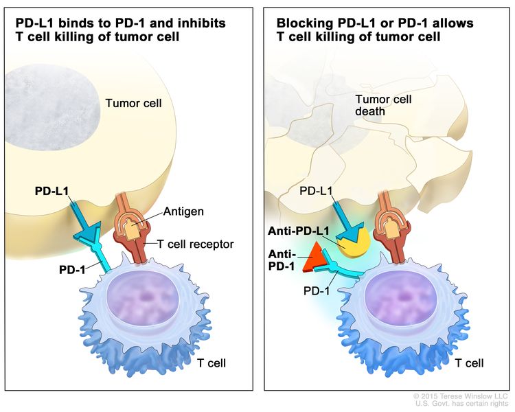
Terapia con inhibidores de PD-1 y PD-L1 : PD-1 es una proteína en la superficie de las células T que ayuda a mantener bajo control las respuestas inmunitarias del cuerpo. La PD-L1 es una proteína que se encuentra en algunos tipos de células cancerosas. Cuando la PD-1 se une a la PD-L1, impide que la célula T mate a la célula cancerosa. Los inhibidores de PD-1 y PD-L1 evitan que las proteínas PD-1 y PD-L1 se unan entre sí. Esto permite que las células T eliminen las células cancerosas.
Inhibidor de puestos de control inmunitario. Las proteínas de puestos de control, como PD-L1 en las células tumorales y PD-1 en las células T, regulan las respuestas inmunitarias. La unión de PD-L1 y PD-1 evita que las células T destruyan las células tumorales en el cuerpo (panel izquierdo). El bloqueo de esta unión con un inhibidor del puesto de control inmunitario (anti-PD-L1 o anti-PD-1) permite que las células T eliminen las células tumorales (panel derecho).
¿Cómo actúan los anticuerpos monoclonales para tratar el cáncer? En este video se explica cómo los anticuerpos monoclonales, como el trastuzumab, el pembrolizumab y el rituximab, bloquean moléculas que las células cancerosas necesitan para multiplicarse, marcan células cancerosas para que el sistema inmunitario las destruya o transportan sustancias que dañan estas células.
Los pacientes pueden evaluar la posibilidad de participar en un ensayo clínico.
Para algunos pacientes, participar en un ensayo clínico puede ser la mejor opción de tratamiento. Los ensayos clínicos son parte del proceso de investigación del cáncer y se realizan para determinar si los nuevos tratamientos para el cáncer son seguros y eficaces o mejores que el tratamiento estándar.
Muchos de los tratamientos estándar actuales para el cáncer se basan en ensayos clínicos anteriores. Los pacientes que participan en un ensayo clínico pueden recibir el tratamiento estándar o ser de los primeros en recibir uno nuevo.
Los pacientes que participan en ensayos clínicos también ayudan a mejorar la forma en que se tratará el cáncer en el futuro. Incluso cuando los ensayos clínicos no conducen a nuevos tratamientos efectivos, suelen responder a preguntas clave y contribuir de forma significativa al avance de la investigación.
Los pacientes pueden participar en ensayos clínicos antes, durante o después de comenzar el tratamiento contra el cáncer.
Algunos ensayos clínicos solo incluyen a pacientes que aún no han recibido tratamiento. Otros ensayos prueban tratamientos para pacientes cuyo cáncer no ha mejorado. También hay ensayos clínicos que prueban nuevas formas de evitar que el cáncer recidive (regrese) o de reducir los efectos secundarios del tratamiento del cáncer.
Se están realizando ensayos clínicos en muchas partes del país. Puede encontrar información sobre los ensayos clínicos respaldados por el NCI en el sitio web de búsqueda de ensayos clínicos del NCI. Puede encontrar ensayos clínicos respaldados por otras organizaciones en el sitio web ClinicalTrials.gov.
Pueden ser necesarias pruebas de seguimiento.
A medida que avanza el tratamiento, se le realizarán pruebas o controles de seguimiento. Es posible que se repitan algunas pruebas para diagnosticar o estadificar el cáncer con el fin de evaluar cómo está funcionando el tratamiento. Las decisiones sobre si continuar, modificar o suspender el tratamiento pueden basarse en los resultados de estas pruebas.
Algunas pruebas seguirán realizándose de manera periódica después de terminar el tratamiento. Los resultados pueden indicar si su afección ha cambiado o si el cáncer ha redicivado (regresado).
Tratamiento del sarcoma de tejido blando en estadio I
Puede utilizar la búsqueda de ensayos clínicos y encontrar ensayos clínicos sobre cáncer patrocinados por el NCI que acepten participantes. La búsqueda le permite filtrar los ensayos según el tipo de cáncer, la edad y el lugar donde se realizan los ensayos. También encontrará información general sobre los ensayos clínicos.
Tratamiento del sarcoma de tejido blando en estadio II y el sarcoma de tejido blando en estadio III que no se diseminó a los ganglios linfáticos
El tratamiento del sarcoma de tejido blando en estadio II y del sarcoma de tejido blando en estadio III que no se ha extendido a los ganglios linfáticos puede incluir las siguientes opciones:
Puede utilizar la búsqueda de ensayos clínicos y encontrar ensayos clínicos sobre cáncer patrocinados por el NCI que acepten participantes. La búsqueda le permite filtrar los ensayos según el tipo de cáncer, la edad y el lugar donde se realizan los ensayos. También encontrará información general sobre los ensayos clínicos.
Tratamiento del sarcoma de tejido blando en estadio III que se ha extendido a los ganglios linfáticos (avanzado)
El tratamiento del sarcoma de tejido blando en estadio III que se ha extendido a los ganglios linfáticos (avanzado) puede incluir las siguientes opciones:
Puede utilizar la búsqueda de ensayos clínicos y encontrar ensayos clínicos sobre cáncer patrocinados por el NCI que acepten participantes. La búsqueda le permite filtrar los ensayos según el tipo de cáncer, la edad y el lugar donde se realizan los ensayos. También encontrará información general sobre los ensayos clínicos.
Tratamiento del sarcoma de tejido blando en estadio IV
Puede utilizar la búsqueda de ensayos clínicos y encontrar ensayos clínicos sobre cáncer patrocinados por el NCI que acepten participantes. La búsqueda le permite filtrar los ensayos según el tipo de cáncer, la edad y el lugar donde se realizan los ensayos. También encontrará información general sobre los ensayos clínicos.
Tratamiento del sarcoma de tejido blando recidivante
Puede utilizar la búsqueda de ensayos clínicos y encontrar ensayos clínicos sobre cáncer patrocinados por el NCI que acepten participantes. La búsqueda le permite filtrar los ensayos según el tipo de cáncer, la edad y el lugar donde se realizan los ensayos. También encontrará información general sobre los ensayos clínicos.
Más información sobre el sarcoma de tejidos blandos
Para obtener más información del National Cancer Institute sobre los sarcomas de tejidos blandos, consulte los siguientes enlaces:
El Physician Data Query (PDQ) es la base de datos integral sobre el cáncer del National Cancer Institute (NCI). La base de datos del PDQ contiene resúmenes con la última información publicada sobre prevención, detección, genética, tratamiento, atención médica de apoyo y medicina complementaria y alternativa relacionada con el cáncer. La mayoría de los resúmenes se presentan en dos versiones. Las versiones para profesionales de la salud contienen información detallada escrita en lenguaje técnico. Las versiones para pacientes están escritas en un lenguaje fácil de entender y no tan técnico. Ambas versiones contienen información precisa y actualizada sobre el cáncer. La mayoría de las versiones también están disponibles en español.
El PDQ es un servicio del NCI. El NCI es parte de los Institutos Nacionales de Salud (NIH), que son el centro de investigación biomédica del Gobierno federal. Los resúmenes del PDQ se basan en una revisión independiente de la literatura médica. No son declaraciones de políticas del NCI ni de los NIH.
Propósito de este resumen
Este resumen de información sobre el cáncer del PDQ contiene información actualizada sobre el tratamiento del sarcoma de tejido blando. Su objetivo es informar y ayudar a los pacientes, las familias y los cuidadores. No proporciona directrices ni recomendaciones formales para tomar decisiones sobre la atención médica.
Revisores y actualizaciones
Los comités editoriales escriben los resúmenes de información sobre el cáncer del PDQ y los mantienen actualizados. Estos comités están formados por equipos de especialistas en el tratamiento del cáncer y otras especialidades relacionadas con esta enfermedad. Los resúmenes se revisan periódicamente y se modifican cuando hay información nueva. La fecha de actualización al pie de cada resumen indica cuándo se realizó el cambio más reciente.
Un ensayo clínico es un estudio para responder a una pregunta científica como, por ejemplo, si un tratamiento es mejor que otro. Los ensayos se basan en estudios anteriores y en lo aprendido en el laboratorio. Cada ensayo responde a determinadas preguntas científicas que permiten encontrar nuevas y mejores formas de ayudar a los pacientes con cáncer. Durante los ensayos clínicos de tratamiento, se recopila información sobre los efectos de un nuevo tratamiento y su eficacia. Si un ensayo clínico demuestra que un nuevo tratamiento es mejor que uno que se utiliza actualmente, el nuevo tratamiento puede convertirse en “estándar”. Los pacientes pueden valorar la posibilidad de participar en un ensayo clínico. Algunos ensayos clínicos solo están abiertos a pacientes que no hayan iniciado el tratamiento.
Los ensayos clínicos se pueden encontrar en línea en el sitio web del NCI. Para obtener más información, llame al Servicio de Información sobre el Cáncer (CIS, por sus siglas en inglés), el centro de contacto del NCI, al 1-800-4-CANCER (1-800-422-6237).
Permiso de uso de este resumen
Physician Data Query (PDQ) es una marca registrada. Se autoriza el libre uso del contenido de los documentos del PDQ como texto. Sin embargo, no se podrá identificar como un resumen de información sobre cáncer del PDQ del NCI, salvo que se reproduzca en su totalidad y se actualice con regularidad. Por otra parte, se permite que los autores incluyan una oración como “en el resumen del PDQ del NCI sobre la prevención del cáncer de mama se describen, de manera concisa, los siguientes riesgos: [incluir fragmento del resumen]”.
La forma recomendada para citar este resumen del PDQ es:
Las imágenes de este resumen se utilizan con el permiso del autor, artista y/o editorial para uso exclusivo en los resúmenes del PDQ. Si desea usar una imagen de un resumen del PDQ sin incluir el resumen completo, debe obtener autorización del propietario. El National Cancer Institute no puede otorgar dicho permiso. Para obtener más información sobre el uso de las imágenes de este resumen o de otras ilustraciones relacionadas con el cáncer, consulte Visuals Online, una colección de más de 3,000 imágenes científicas.
Descargo de responsabilidad
La información de estos resúmenes no debe utilizarse para tomar decisiones sobre reembolsos de seguros. Puede encontrar más información sobre la cobertura de seguros en Cancer.gov en el sitio Manejo de la atención del cáncer.
Contáctenos
Puede encontrar más información sobre cómo contactarnos o recibir ayuda en el sitio web Cancer.gov en la página Comuníquese con el NCI. También puede enviar sus preguntas a Cancer.gov en el apartado Escríbanos del sitio web.
Actualizado:
Este contenido ha sido facilitado por el National Cancer Institute (www.cancer.gov)