


Tratamiento del cáncer de mama en mujeres
Obtenga una atención excepcional para el cáncer de mama, uno de los cánceres más comunes que afectan a las mujeres, en el Montefiore Einstein Comprehensive Cancer Center. Como uno de los primeros centros del cáncer designados por el NCI durante más de 50 años, hemos sido líderes en la investigación, el diagnóstico y el tratamiento de más de 200 tipos de cáncer.
Espere una atención integral y personalizada cáncer de mama que cumpla con los más altos estándares de calidad y seguridad. El Montefiore Einstein Comprehensive Cancer Center está clasificado dentro del 1 % de los mejores hospitales del país en cuanto a atención médica del cáncer según U.S. News & World Report . Nuestro programa de cáncer de mama también está acreditado por el Programa Nacional de Acreditación para Centros de Cáncer de Mama (NAPBC), siguiendo los más altos estándares de calidad para la atención del cáncer de mama .
En un entorno moderno y tranquilo diseñado para promover la curación, nuestro programa de cáncer de mama:
- Ofrece las últimas y más efectivas pruebas de diagnóstico, tratamientos y tecnología avanzada, entre ellas:
- Pruebas de diagnóstico en el mismo día para mamografía utilizando tomosíntesis 3D de última generación, ecografía y biopsia con aguja mínimamente invasiva
- Navegación quirúrgica para la cirugía de conservación de mama, lo que nos convierte en uno de los pocos programas de cáncer de mama en EE. UU. que ofrece esta opción, que le permite alcanzar el mejor resultado en la menor cantidad de cirugías posible.
- Técnicas de radioterapia, como tratamientos de radiación de dosis única durante la cirugía, para tratar el cáncer más rápidamente que la radioterapia tradicional
- Técnicas de lumpectomía que usan tecnología de imágenes durante la cirugía para extirpar completamente el cáncer en el 90 % de los pacientes, una cifra significativamente superior al promedio nacional del 65 % en otros programas.
- Le da acceso a nuevos tratamientos prometedores a través de ensayos clínicos y trabaja activamente para aumentar la accesibilidad a los ensayos clínicos y eliminar las barreras de acceso a la atención para todas las personas.
- Pioneros en avances en la prevención, diagnóstico y tratamiento del cáncer de mama a través de investigaciones de vanguardia, como el estudio histórico TAILORx, que descubrió que hasta el 70% de las mujeres con cáncer de mama en etapa temprana pueden evitar la quimioterapia mediante la terapia hormonal, y otros descubrimientos científicos que están dando forma al futuro de la atención del cáncer de mama.
- Pone a disposición medidas de prevención y terapias para el linfedema reconocidas a nivel mundial, como el enfoque curativo preventivo microquirúrgico linfático (LYMPHA), que se desarrolló en nuestro centro del cáncer.
- Se centra en su bienestar integral durante y después del tratamiento del cáncer a través de atención, programas y recursos integrales y de apoyo.
- Le permite tomar decisiones informadas sobre su atención a través del apoyo y la educación.
- Está dirigido por un equipo multidisciplinario de especialistas en cáncer de mama, compuesto por oncólogos médicos, oncólogos quirúrgicos, cirujanos plásticos y reconstructivos, radioterapeutas, trabajadores sociales y otros profesionales dedicados, que trabajan juntos para garantizar que reciba una atención excepcional, coordinada y centrada en el paciente y le ofrece citas combinadas el mismo día para su comodidad.
Cuando necesite atención para el cáncer de mama, recurra a nuestros proveedores que se apasionan por acabar con el cáncer y abordar todas sus necesidades de salud.
Acuda al Centro de Salud Mamaria para recibir una atención médica coordinada y especializada en un entorno moderno, acceder a algunos de los mejores especialistas multidisciplinarios del país y contar con toda la información y orientación personalizada que necesita para dar los próximos pasos.

El Montefiore Einstein Comprehensive Cancer Center, designado como centro integral del cáncer por el National Cancer Institute (NCI), apoya la misión y las normas del NCI. La siguiente información sobre los tipos de cáncer, prevención y tratamientos ha sido facilitada por el NCI.
Tratamiento del cáncer de mama (PDQ®): versión para pacientes
Información general sobre el cáncer de mama
Puntos clave
- El cáncer de mama es una enfermedad en la que se forman células malignas (cancerosas) en los tejidos de la mama.
- Un historial familiar de cáncer de mama y otros factores aumentan el riesgo de desarrollarlo.
- El cáncer de mama a veces está causado por mutaciones (cambios) genéticos hereditarios.
- El uso de ciertos medicamentos y otros factores disminuyen el riesgo de desarrollar cáncer de mama.
- Entre los signos del cáncer de mama se encuentran un bulto o algún cambio en la mama.
- Para diagnosticar el cáncer de mama se utilizan pruebas que examinan los senos.
- Si se detecta cáncer, se realizan pruebas para estudiar las células cancerosas.
- Certain factors affect prognosis (chance of recovery) and treatment options.
El cáncer de mama es una enfermedad en la que se forman células malignas (cancerosas) en los tejidos de la mama.
La mama está formada por lóbulos y conductos. Cada mama tiene de 15 a 20 secciones llamadas lóbulos, los cuales, a su vez, se dividen en secciones más pequeñas llamadas lobulillos. Estos terminan en docenas de pequeños bulbos que pueden producir leche. Los lóbulos, lobulillos y bulbos están conectados entre sí por tubos delgados llamados conductos.
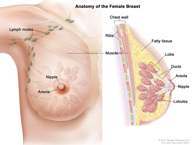
La mama femenina contiene lóbulos, lobulillos y conductos que producen y transportan leche hasta el pezón. El tejido adiposo le da forma, mientras que los músculos y la pared torácica la sostienen. El sistema linfático, incluidos los ganglios linfáticos, filtra la linfa y almacena glóbulos blancos que ayudan a combatir infecciones y enfermedades.
Cada seno también tiene vasos sanguíneos y vasos linfáticos. Los vasos linfáticos transportan un líquido acuoso casi incoloro llamado linfa. Los vasos linfáticos transportan linfa entre los ganglios linfáticos. Los ganglios linfáticos son estructuras pequeñas con forma de frijol que se encuentran en todo el cuerpo. Filtran la linfa y almacenan glóbulos blancos que ayudan a combatir infecciones y enfermedades. Se encuentran grupos de ganglios linfáticos cerca del seno en la axila (debajo del brazo), encima de la clavícula y en el pecho.
El tipo más común de cáncer de mama es el carcinoma ductal, que se origina en las células de los conductos y representa entre el 70 % y el 80 % de todos los casos de cáncer de mama. El segundo tipo más común es el carcinoma lobulillar, que se origina en los lóbulos y representa entre el 10 % y el 15 % de todos los casos de cáncer de mama. El carcinoma lobulillar se presenta con mayor frecuencia en ambas mamas simultáneamente que otros tipos de cáncer de mama. El cáncer de mama inflamatorio es un tipo poco común de cáncer de mama de rápido crecimiento en el que las células cancerosas obstruyen los vasos linfáticos de la piel de la mama.
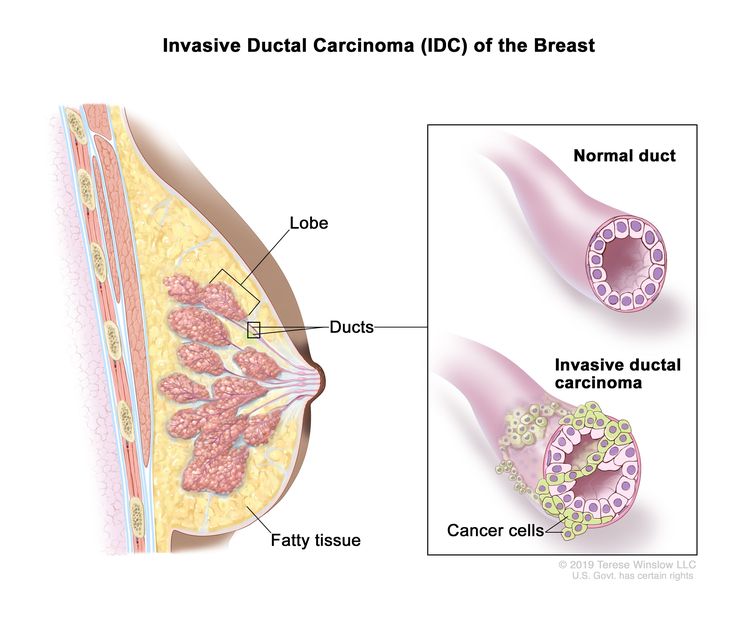
El carcinoma ductal invasivo (CDI) de mama se origina en el revestimiento de un conducto mamario (conducto galactóforo) y se propaga a otros tejidos de la mama. También puede diseminarse a través del sistema sanguíneo y linfático a otras partes del cuerpo. El CDI es el tipo más común de cáncer de mama invasivo.
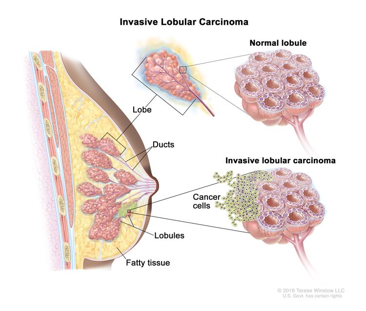
El carcinoma lobulillar invasivo comienza en los lobulillos (glándulas mamarias) de la mama y se propaga a otros tejidos de la mama. También puede diseminarse a través de los sistemas sanguíneo y linfático a otras partes del cuerpo.
Para obtener más información sobre el cáncer de mama, consulte los siguientes sitios web:
Un historial familiar de cáncer de mama y otros factores aumentan el riesgo de desarrollarlo.
Todo lo que aumenta la probabilidad de padecer una enfermedad se denomina factor de riesgo. Tener un factor de riesgo no significa que padecerá cáncer; no tener factores de riesgo no significa que no padecerá cáncer. Hable con su médico si cree que puede estar en riesgo de padecer cáncer de mama.
Los factores de riesgo del cáncer de mama son los siguientes:
- Historial personal de cáncer de mama invasivo, carcinoma ductal in situ (CDIS) o carcinoma lobulillar in situ (CLIS)
- Historial personal de enfermedad mamaria benigna (no cancerosa)
- Historial familiar de cáncer de mama en un familiar de primer grado (madre, hija o hermana)
- Cambios hereditarios en los genes BRCA1 o BRCA2 o en otros genes que aumentan el riesgo de desarrollar cáncer de mama.
- Tejido mamario que es denso en una mamografía
- Exposición del tejido mamario al estrógeno producido por el cuerpo. Esto puede estar causado por:
- Haber tenido la menstruación a una edad temprana.
- Edad avanzada al momento del primer parto o no haber tenido hijos
- Inicio de la menopausia a una edad más avanzada
- Tomar hormonas como el estrógeno combinado con progestina para los síntomas de la menopausia
- Tratamiento con radioterapia en la mama o tórax
- Consumo de alcohol
- Obesidad.
La edad avanzada es el principal factor de riesgo para la mayoría de los cánceres. La probabilidad de padecer cáncer aumenta con la edad.
La herramienta de evaluación del riesgo de cáncer de mama del NCI utiliza los factores de riesgo de una mujer para estimar su riesgo de desarrollar cáncer de mama durante los próximos cinco años y hasta los 90 años. Esta herramienta en línea está diseñada para ser utilizada por un proveedor de atención médica. Para obtener más información sobre el riesgo de cáncer de mama, llame al 1-800-4-CANCER.
El cáncer de mama a veces está causado por mutaciones (cambios) genéticos hereditarios.
Los genes de las células contienen la información hereditaria que se recibe de los padres de una persona. El cáncer de mama hereditario representa aproximadamente entre el 5 % y el 10% de todos los cáncer de mama. Algunos genes mutados relacionados con el cáncer de mama son más comunes en ciertos grupos étnicos.
Las mujeres que tienen ciertas mutaciones genéticas, como una mutación BRCA1 o BRCA2, tienen un mayor riesgo de cáncer de mama. Estas mujeres también tienen un mayor riesgo de cáncer de ovario y pueden tener un mayor riesgo de otros tipos de cáncer. Los hombres que tienen un gen mutado relacionado con el cáncer de mama también tienen un mayor riesgo de cáncer de mama. Para obtener más información, consulte Tratamiento del cáncer de mama masculino.
Existen pruebas que pueden detectar (encontrar) genes mutados. Estas pruebas genéticas a veces se realizan a miembros de familias con alto riesgo de cáncer. Para obtener más información, consulte Genética de los cánceres de mama y ginecológicos.
El uso de ciertos medicamentos y otros factores disminuyen el riesgo de desarrollar cáncer de mama.
Cualquier circunstancia que disminuya la probabilidad de desarrollar una enfermedad se llama factor de protección.
Los factores de protección contra el cáncer de mama incluyen los siguientes:
- Tomar alguno de los siguientes:
- Menor exposición del mama tejidos hacia el estrógeno hecho por el cuerpo. Esto puede ser resultado de:
- Embarazo temprano
- Amamantamiento
- Hacer suficiente ejercicio
- Haberse realizado alguno de los siguientes procedimientos:
- Mastectomía para reducir el riesgo de cáncer
- Ooforectomía para reducir el riesgo de cáncer
- Ablación ovárica.
Entre los signos del cáncer de mama se encuentran un bulto o algún cambio en la mama.
Estos y otros síntomas pueden ser causados por cáncer de mama u otras afecciones. Consulte a su médico si presenta alguno de los siguientes síntomas:
- Un bulto o engrosamiento en la mama o cerca de esta o en el área de la axila
- Un cambio en el tamaño o la forma de la mama
- Un hoyuelo o arrugas en la piel de la mama
- Un pezón invertido hacia el interior de la mama
- Liberación de un líquido, que no es leche materna, del pezón, especialmente si tiene sangre.
- Piel escamosa, enrojecida o hinchada en la mama, el pezón o la areola (el área oscura de la piel alrededor del pezón)
- Hoyuelos en la mama que hacen que la piel se vea como la de una naranja, lo que se conoce como peau d’orange.
Para diagnosticar el cáncer de mama se utilizan pruebas que examinan los senos.
Consulte con su médico si nota algún cambio en sus mamas. Se pueden utilizar las siguientes pruebas y procedimientos:
- Reconocimiento físico e historial de salud: un examen del cuerpo para evaluar el estado general de salud, incluida la detección de signos de enfermedad, como bultos o cualquier otra anomalía. También se toma nota de los hábitos de salud del paciente y de sus enfermedades y tratamientos previos.
- Examen clínico de senos (ECS): es un examen de los senos realizado por un médico u otro profesional de la salud. El médico palpará cuidadosamente las mamas y las axilas para detectar bultos o cualquier anomalía.
- Mamografía: un rayos X. de las mamas.
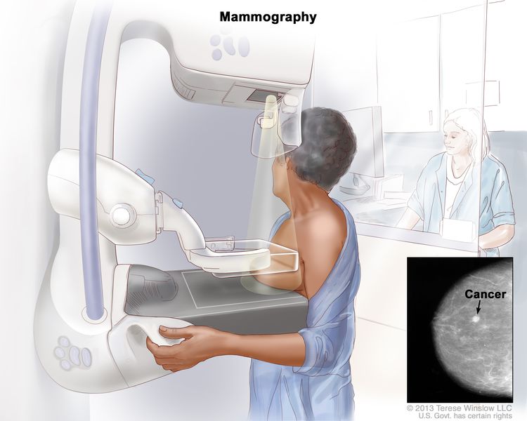
La mamografía es una prueba de imagen que se utiliza para detectar y diagnosticar el cáncer de mama. Puede detectar tejido mamario anómalo, incluido el cáncer, a veces incluso antes de que aparezcan los síntomas.
- Exploración por ecografía: es un procedimiento en el que ondas sonoras de alta energía (ultrasonido) rebotan en tejidos u órganos internos y producen ecos. Los ecos forman una imagen de los tejidos del cuerpo llamada ecograma. La imagen se puede imprimir para su posterior análisis.
- MRI (imagen por resonancia magnética): procedimiento que utiliza un imán, ondas de radio, y una computadora para hacer una serie de imágenes detalladas de ambos senos. Este procedimiento también se llama resonancia magnética nuclear (RMN).
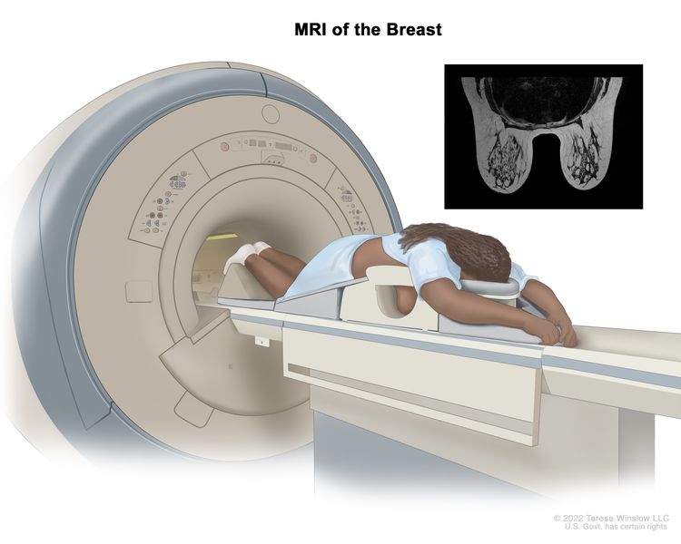
La resonancia magnética (RM) de mama es un procedimiento que utiliza ondas de radio, un potente imán y una computadora para crear imágenes detalladas del interior de la mama. Se puede inyectar un medio de contraste en una vena (no se muestra) para facilitar la visualización de los tejidos mamarios en las imágenes de RM. La RM se puede utilizar junto con otras pruebas de imagen mamaria para detectar cáncer de mama u otros cambios anormales en la mama. También se puede utilizar para la detección precoz del cáncer de mama en personas con alto riesgo de padecer la enfermedad. Nota: La imagen insertada muestra una resonancia magnética del interior de ambas mamas. Crédito de la imagen insertada: The Cancer Imaging Archive (TCIA).
- Blood chemistry studies: A procedure in which a blood sample is checked to measure the amounts of certain substances released into the blood by organs and tissues in the body. An unusual (higher or lower than normal) amount of a substance can be a sign of disease.
- Biopsy: extracción de células o tejidos para que puedan ser observados con un microscopio por un pathologist para detectar signos de cáncer. Si se encuentra un bulto en la mama, se puede realizar una biopsia.
Hay cuatro tipos de biopsia que se utilizan para detectar el cáncer de mama:
- Biopsia escisional: extirpación de un bulto completo de tejido
- Biopsia incisional: extracción de parte de un bulto o de una muestra de tejido
- Biopsia central: extracción de tejido mediante una aguja ancha
- Biopsia por aspiración con aguja fina (PAAF): extracción de tejido o líquido mediante una aguja fina
Si se detecta cáncer, se realizan pruebas para estudiar las células cancerosas.
Las decisiones sobre el tratamiento más adecuado se basan en los resultados de estas pruebas. Las pruebas ofrecen información sobre:
- Qué tan rápido puede crecer el cáncer.
- La probabilidad de que el cáncer se propague por el cuerpo.
- Qué tan bien podrían funcionar ciertos tratamientos.
- La probabilidad de que el cáncer recidive (regrese).
Las pruebas incluyen:
- Prueba de receptores de estrógeno y progesterona: es una prueba para medir la cantidad de receptores de estrógeno y progesterona (hormonas) en el tejido canceroso. Si hay más receptores de estrógeno y progesterona de lo normal, el cáncer se denomina positivo para receptores de estrógeno y/o progesterona. Este tipo de cáncer de mama puede crecer más rápidamente. Los resultados de la prueba muestran si el tratamiento para bloquear el estrógeno y la progesterona puede detener el crecimiento del cáncer.
- Prueba del receptor 2 del factor de crecimiento epidérmico humano (HER2/neu): Una prueba de laboratorio para medir cuántos genes HER2/neu hay y cuánta proteína HER2/neu se produce en una muestra de tejido. Si hay más genes HER2/neu o niveles más altos de proteína HER2/neu de lo normal, el cáncer se denomina HER2/neu positivo o HER2 positivo. Este tipo de cáncer de mama puede crecer más rápidamente y tiene más probabilidades de diseminarse a otras partes del cuerpo. El cáncer puede tratarse con medicamentos que actúan sobre la proteína HER2/neu, como trastuzumab y pertuzumab .
- Pruebas multigénicas: son pruebas en las que se estudian muestras de tejido para observar la actividad de muchos genes al mismo tiempo. Pueden ayudar a predecir si el cáncer se propagará a otras partes del cuerpo o si recidivará (regresará).
Existen muchos tipos de pruebas multigénicas. Se han estudiado en ensayos clínicos las siguientes pruebas multigénicas:
- Oncotipo DX: esta prueba ayuda a predecir si el cáncer de mama en etapa temprana, que es positivo para el receptor de estrógeno y negativo para los ganglios linfáticos, se propagará a otras partes del cuerpo. Si el riesgo de que el cáncer se propague es alto, se puede administrar quimioterapia para reducir el riesgo.
- MammaImprimir: es un análisis de laboratorio en el que se analiza la actividad de 70 genes diferentes en el tejido del cáncer de mama de mujeres que tienen cáncer de mama invasivo en estadio temprano que no se ha propagado a los ganglios linfáticos o que lo ha hecho a tres ganglios linfáticos o menos. El nivel de actividad de estos genes ayuda a predecir si el cáncer se propagará a otras partes del cuerpo o regresará. Si el análisis muestra que el riesgo de que se propague o regrese es alto, se puede administrar quimioterapia para reducir el riesgo.
Según estas pruebas, el cáncer de mama podría describirse como uno de los siguientes tipos:
- Receptor hormonal positivo (positivo para receptores de estrógeno y/o progesterona) o receptor hormonal negativo (negativo para receptores de estrógeno y/o progesterona)
- Positivo para HER2 o negativo para HER2
- Triple negativo (negativo para receptores de estrógeno, receptores de progesterona y HER2)
Esta información ayuda al médico a decidir qué tratamientos funcionarán mejor para su tipo de cáncer.
Certain factors affect prognosis (chance of recovery) and treatment options.
The prognosis and treatment options depend on:
- El estadio del cáncer (el tamaño del tumor y si está solo en la mama o se ha diseminado a los ganglios linfáticos u otras partes del cuerpo).
- El tipo de cáncer de mama
- Niveles del receptor de estrógeno y del receptor de progesterona en el tejido tumoral
- Niveles del receptor del factor de crecimiento epidérmico humano tipo 2 (HER2/neu) en el tejido tumoral
- Si el tejido tumoral es triple negativo (células que no tienen receptores de estrógeno, receptores de progesterona o niveles altos de HER2/neu).
- La rapidez con la que está creciendo el tumor
- La probabilidad de que el tumor recidive (regrese).
- La edad, la salud general y el estado menopáusico de una mujer (si todavía tiene periodos menstruales.
- Si el cáncer se acaba de diagnosticar o recidivó (volvió).
Estadios del cáncer de mama
Puntos clave
- Después de diagnosticar el cáncer de mama, se realizan pruebas para determinar si las células cancerosas se han diseminado dentro de la mama o a otras partes del cuerpo.
- There are three ways that cancer spreads in the body.
- El cáncer puede extenderse desde donde comenzó a otras partes del cuerpo.
- En el cáncer de mama, el estadio se basa en el tamaño y la ubicación del tumor primario, la propagación del cáncer a los ganglios linfáticos cercanos u otras partes del cuerpo, el grado del tumor y la presencia de ciertos biomarcadores.
- El sistema TNM se usa para describir el tamaño del tumor primario y la diseminación del cáncer a los ganglios linfáticos cercanos u otras partes del cuerpo.
- Tumor (T): el tamaño y la ubicación del tumor
- Nódulos linfáticos (N): el tamaño y la ubicación de los ganglios linfáticos a los que se ha propagado el cáncer.
- Metástasis (M): la propagación del cáncer a otras partes del cuerpo
- El sistema de gradación se usa para describir qué tan rápido es probable que un tumor de mama crezca y se propague.
- La prueba de biomarcadores se utiliza para determinar si las células del cáncer de mama tienen ciertos receptores.
- El sistema TNM, el sistema de gradación y el estado de los biomarcadores se combinan para determinar el estadio del cáncer de mama.
- Hable con su médico para saber cuál es su estadio de cáncer de mama y cómo se utiliza para planificar el tratamiento más adecuado para usted.
- El tratamiento del cáncer de mama depende, en parte, del estadio de la enfermedad.
Después de diagnosticar el cáncer de mama, se realizan pruebas para determinar si las células cancerosas se han diseminado dentro de la mama o a otras partes del cuerpo.
El proceso que se utiliza para determinar si el cáncer se ha diseminado dentro de la mama o a otras partes del cuerpo se denomina estadificación . La información obtenida durante la estadificación determina la etapa de la enfermedad. Es importante conocer la etapa para planificar el tratamiento. Los resultados de algunas de las pruebas utilizadas para diagnosticar el cáncer de mama también se utilizan para estadificar la enfermedad. (Véase la sección de Información general ).
También se pueden utilizar las siguientes pruebas y procedimientos en el proceso de estadificación:
- Biopsia del ganglio linfático centinela: Extirpación del ganglio linfático centinela durante la cirugía. El ganglio linfático centinela es el primer ganglio linfático de un grupo que recibe el drenaje linfático del tumor primario . Es el primer ganglio linfático al que es probable que se disemine el cáncer desde el tumor primario. Se inyecta una sustancia radiactiva y/o un tinte azul cerca del tumor. La sustancia o el tinte fluye a través de los conductos linfáticos hacia los ganglios linfáticos. Se extirpa el primer ganglio linfático que recibe la sustancia o el tinte. Un patólogo examina el tejido al microscopio para buscar células cancerosas. Si no se encuentran células cancerosas, puede que no sea necesario extirpar más ganglios linfáticos. A veces, se encuentra un ganglio linfático centinela en más de un grupo de ganglios.
- Radiografía de tórax: radiografía de los órganos y huesos del interior del tórax. Un rayo X es un tipo de haz de energía que puede atravesar el cuerpo y plasmarse en una película, generando una imagen de áreas internas del cuerpo.
- Tomografía computarizada (TC): Procedimiento que genera una serie de imágenes detalladas de áreas internas del cuerpo, tomadas desde diferentes ángulos. Las imágenes se obtienen mediante una computadora conectada a un equipo de rayos X. Se puede inyectar un medio de contraste en una vena o ingerirlo para que los órganos o tejidos se visualicen con mayor claridad. Este procedimiento también se conoce como tomografía computarizada o tomografía axial computarizada.
- Exploración ósea: es un procedimiento para comprobar si hay células que se dividen rápidamente, como células cancerosas, en el hueso. Se inyecta una cantidad muy pequeña de material radiactivo en una vena y viaja a través del torrente sanguíneo. El material radiactivo se acumula en los huesos con cáncer y es detectado por un escáner.
- Exploración PET (tomografía por emisión de positrones): es un procedimiento para detectar células tumorales malignas en el cuerpo. Se inyecta una pequeña cantidad de glucosa radiactiva (azúcar) en una vena. El escáner PET gira alrededor del cuerpo y genera una imagen de las zonas donde se utiliza la glucosa. Las células tumorales malignas se ven más brillantes en la imagen porque presentan un mayor nivel de actividad y absorben más glucosa que las células normales.
There are three ways that cancer spreads in the body.
Cancer can spread through tissue, the lymph system, and the blood:
- Tissue. The cancer spreads from where it began by growing into nearby areas.
- Lymph system. The cancer spreads from where it began by getting into the lymph system. The cancer travels through the lymph vessels to other parts of the body.
- Blood. The cancer spreads from where it began by getting into the blood. The cancer travels through the blood vessels to other parts of the body.
El cáncer puede extenderse desde donde comenzó a otras partes del cuerpo.
Cuando el cáncer se extiende a otra parte del cuerpo se denomina metástasis. Las células cancerosas se desprenden de donde comenzaron (tumor primario) y viajan a través del sistema linfático o la sangre.
- Sistema linfático: el cáncer entra en el sistema linfático, viaja a través de los vasos linfáticos y forma un tumor (tumor metastásico) en otra parte del cuerpo.
- Sangre: el cáncer llega a la sangre, viaja a través de los vasos sanguíneos y forma un tumor (tumor metastásico) en otra parte del cuerpo.
El tumor metastásico es el mismo tipo de cáncer que el tumor primario. Por ejemplo, si el cáncer de mama se propaga al hueso, las células cancerosas del hueso son en realidad células de cáncer de mama . La enfermedad es cáncer de mama metastásico, no cáncer de hueso .
En el cáncer de mama, el estadio se basa en el tamaño y la ubicación del tumor primario, la propagación del cáncer a los ganglios linfáticos cercanos u otras partes del cuerpo, el grado del tumor y la presencia de ciertos biomarcadores.
Para planificar el mejor tratamiento posible y comprender su pronóstico, es importante conocer el estadio del cáncer de mama.
Existen tres tipos de grupos de estadios del cáncer de mama:
- El estadio pronóstico clínico se utiliza inicialmente para asignar un estadio a todos los pacientes según su historial de salud, un reconocimiento físico, las pruebas de diagnóstico por imágenes (si se realizan) y las biopsias. El estadio pronóstico clínico se describe mediante el sistema TNM, el grado del tumor y el estado de los biomarcadores (ER, PR, HER2). En la estadificación clínica, se utiliza la mamografía o la ecografía para examinar los ganglios linfáticos en busca de signos de cáncer.
- El estadio pronóstico patológico se utiliza luego para pacientes que se realicen una cirugía como primer tratamiento. El estadio pronóstico patológico se basa en toda la información clínica, el estado de los biomarcadores y los resultados de las pruebas de laboratorio del tejido mamario y los ganglios linfáticos extirpados durante la cirugía.
- El estadio anatómico se basa en el tamaño y la propagación del cáncer según lo describe el sistema TNM. El estadio anatómico se utiliza en partes del mundo donde las pruebas de biomarcadores no están disponibles. No se utiliza en Estados Unidos.
El sistema TNM se usa para describir el tamaño del tumor primario y la diseminación del cáncer a los ganglios linfáticos cercanos u otras partes del cuerpo.
En el caso del cáncer de mama, el sistema TNM describe el tumor de la siguiente manera:
Tumor (T): el tamaño y la ubicación del tumor
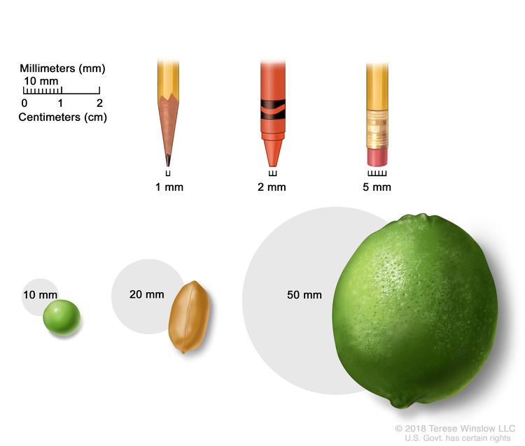
A menudo, el tamaño de los tumores se miden en milímetros (mm) o centímetros. Los elementos comunes que se pueden usar para mostrar el tamaño del tumor en mm son: una punta de lápiz afilada (1 mm), una punta de un crayón nuevo (2 mm), un borrador de lápiz (5 mm), un guisante (10 mm), un maní (20 mm) y una lima (50 mm).
- TX: no se puede evaluar el tumor primario.
- T0: no hay signos de un tumor primario en la mama.
- Tis: carcinoma in situ: hay dos tipos de carcinoma de mama in situ:
- Tis ( CDIS ): El CDIS es una afección en la que se encuentran células anormales en el revestimiento del conducto mamario . Estas células anormales no se han propagado fuera del conducto a otros tejidos de la mama. En algunos casos, el CDIS puede convertirse en un cáncer de mama invasivo capaz de propagarse a otros tejidos. Actualmente, no se sabe con certeza qué lesiones pueden volverse invasivas.
- Tis (enfermedad de Paget): la enfermedad de Paget del pezón es una afección en la que hay presencia de células anómalas en las células de la piel del pezón y pueden propagarse a la areola. No se clasifica según el sistema TNM. Si hay enfermedad de Paget Y cáncer de mama invasivo, se utiliza el sistema TNM para clasificar el cáncer de mama invasivo.
- T1: el tumor mide 20 milímetros o menos. Hay cuatro subtipos de un tumor T1 dependiendo del tamaño del tumor:
- T1mi: el tumor mide 1 milímetro o menos.
- T1a: el tumor mide más de 1 milímetro, pero no más de 5 milímetros.
- T1b: el tumor mide más de 5 milímetros, pero no más de 10 milímetros.
- T1c: el tumor mide más de 10 milímetros, pero no más de 20 milímetros.
- T2: el tumor mide más de 20 milímetros, pero no más de 50 milímetros.
- T3: el tumor mide más de 50 milímetros.
- T4: el tumor se describe como uno de los siguientes:
- T4a: el tumor ha crecido hacia la pared torácica.
- T4b: el tumor ha invadido la piel: se ha formado una úlcera en la superficie de la piel de la mama, se han formado pequeños nódulos tumorales en la misma mama que el tumor primario y/o hay hinchazón de la piel de la mama.
- T4c: el tumor ha crecido hacia la pared torácica y la piel.
- T4d: cáncer de mama inflamatorio: un tercio o más de la piel de la mama está roja e hinchada (lo que se denomina piel de naranja).
Nódulos linfáticos (N): el tamaño y la ubicación de los ganglios linfáticos a los que se ha propagado el cáncer.
Cuando se extirpan los ganglios linfáticos mediante cirugía y un patólogo los estudia bajo un microscopio, se utiliza la estadificación patológica para describirlos. A continuación se describe la estadificación patológica de los ganglios linfáticos.
- NX: no se pueden evaluar los ganglios linfáticos.
- N0: no hay signos de cáncer en los ganglios linfáticos o solo hay pequeños grupos de células cancerosas de no más de 0.2 milímetros en ellos.
- N1: el cáncer se describe como uno de los siguientes:
- N1mi: el cáncer se ha diseminado a los ganglios linfáticos axilares (área de la axila) y mide más de 0.2 milímetros, pero no más de 2 milímetros.
- N1a: el cáncer se diseminó a entre uno y tres ganglios linfáticos axilares y el cáncer en al menos uno de los ganglios linfáticos mide más de 2 milímetros.
- N1b: el cáncer se ha diseminado a los ganglios linfáticos cerca del esternón en el mismo lado del cuerpo que el tumor primario. El cáncer mide más de 0.2 milímetros y se detecta mediante una biopsia del ganglio linfático centinela; no está presente en los ganglios linfáticos axilares.
- N1c: el cáncer se diseminó a entre uno y tres ganglios linfáticos axilares y el cáncer en al menos uno de los ganglios linfáticos mide más de 2 milímetros. También se detecta cáncer en una biopsia de ganglio linfático centinela en los ganglios linfáticos cerca del esternón en el mismo lado del cuerpo que el tumor primario.
- N2: el cáncer se describe como uno de los siguientes:
- N2a: el cáncer se diseminó a entre cuatro y nueve ganglios linfáticos axilares y, en al menos uno de los ganglios linfáticos, mide más de 2 milímetros.
- N2b: el cáncer se ha diseminado a los ganglios linfáticos cerca del esternón y se detecta mediante pruebas de imagen. No se detecta cáncer en los ganglios linfáticos axilares mediante una biopsia del ganglio linfático centinela o una disección de los ganglios linfáticos.
- N3: el cáncer se describe como uno de los siguientes:
- N3a: el cáncer se ha diseminado a diez o más ganglios linfáticos axilares y, en al menos uno de los ganglios linfáticos, mide más de 2 milímetros o se ha diseminado a los ganglios linfáticos debajo de la clavícula.
- N3b: el cáncer se diseminó a entre uno y nueve ganglios linfáticos axilares y, en al menos uno de los ganglios linfáticos, mide más de 2 milímetros. También se diseminó a los ganglios linfáticos cerca del esternón y se detecta mediante pruebas de imagen.
o
El cáncer se diseminó a entre cuatro y nueve ganglios linfáticos axilares y, en al menos uno de los ganglios linfáticos, mide más de 2 milímetros. También se diseminó a los ganglios linfáticos cerca del esternón en el mismo lado del cuerpo que el tumor primario y mide más de 0.2 milímetros; se detecta mediante una biopsia de ganglio linfático centinela.
- N3c: el cáncer se diseminó a los ganglios linfáticos por encima de la clavícula en el mismo lado del cuerpo que el tumor primario.
Cuando los ganglios linfáticos se examinan mediante mamografía o ecografía, se denomina estadificación clínica. La estadificación clínica de los ganglios linfáticos no se describe en esta página.
Metástasis (M): la propagación del cáncer a otras partes del cuerpo
- M0: no hay señales de que el cáncer se haya propagado a otras partes del cuerpo.
- M1: el cáncer se ha propagado a otras partes del cuerpo, con mayor frecuencia a los huesos, los pulmones, el hígado o el cerebro. Si se ha propagado a ganglios linfáticos distantes, el cáncer en los ganglios linfáticos mide más de 0.2 milímetros. Se lo denomina cáncer de mamametastásico.
El sistema de gradación se usa para describir qué tan rápido es probable que un tumor de mama crezca y se propague.
El sistema de gradación describe un tumor basándose en cuán anómalas se ven las células y el tejido canceroso bajo un microscopio y qué tan rápido es probable que las células crezcan y se propaguen. Las células cancerosas de bajo grado se parecen más a las células normales y tienden a crecer y diseminarse más lentamente que las de alto grado. Para describir cuán anómalas son las células y el tejido canceroso, el patólogo evaluará las tres características siguientes:
- Qué proporción del tejido tumoral conserva conductos mamarios normales.
- El tamaño y la forma de los núcleos de las células tumorales
- Cuántas células en división están presentes, lo cual indica la velocidad con que las células del tumor crecen y se dividen.
Para cada característica, el patólogo asigna una puntuación del 1 a 3; una puntuación de 1 significa que las células y el tejido tumoral se parecen más a las células y el tejido normales, mientras que una puntuación de 3 significa que las células y el tejido tienen una apariencia más anómala. Las puntuaciones de cada característica se suman para obtener una puntuación total entre 3 y 9.
Hay tres grados posibles:
- Puntuación total de 3 a 5: G1 (grado bajo o bien diferenciado)
- Puntuación total de 6 a 7: G2 (grado intermedio o moderadamente diferenciado)
- Puntuación total de 8 a 9: G3 (grado alto o poco diferenciado)
La prueba de biomarcadores se utiliza para determinar si las células del cáncer de mama tienen ciertos receptores.
Las células mamarias sanas y algunas células de cáncer de mama tienen receptores (biomarcadores) que se adhieren a las hormonas estrógeno y progesterona. Estas hormonas son necesarias para que las células sanas y algunas células de cáncer de mama crezcan y se dividan. Para verificar la presencia de estos biomarcadores, se extraen muestras de tejido que contienen células de cáncer de mama durante una biopsia o cirugía. Las muestras se analizan en un laboratorio para determinar si las células de cáncer de mama tienen receptores de estrógeno o progesterona.
Otro tipo de receptor (biomarcador) que se encuentra en la superficie de todas las células del cáncer de mama se llama HER2. Los receptores HER2 son necesarios para que las células del cáncer de mama crezcan y se dividan.
Para el cáncer de mama, las pruebas de biomarcadores incluyen:
- Receptor de estrógeno (ER): si las células del cáncer de mama tienen receptores de estrógeno, se las denomina ER positivas (ER+). Si no los tienen, se las denomina ER negativas (ER-).
- Receptor de progesterona (PR): si las células del cáncer de mama tienen receptores de progesterona, se las denomina PR positivas (PR+). Si no los tienen, se las denomina PR negativas (PR-).
- Receptor del factor de crecimiento epidérmico humano tipo 2 (HER2/neu o HER2): si las células del cáncer de mama tienen cantidades mayores de lo normal de receptores HER2 en su superficie, las células cancerosas se denominan HER2 positivas (HER2+). Si las células del cáncer de mama tienen una cantidad normal de HER2 en su superficie, las células cancerosas se denominan HER2 negativas (HER2-). El cáncer de mama HER2+ tiene más probabilidades de crecer y dividirse más rápido que el cáncer de mama HER2-.
En ocasiones, las células del cáncer de mama se describen como triple negativas o triple positivas .
- Triple negativo . Si las células del cáncer de mama no tienen receptores de estrógeno, receptores de progesterona o una cantidad mayor de lo normal de receptores HER2, las células cancerosas se denominan triple negativas.
- Triple positivo . Si las células de cáncer de mama tienen receptores de estrógeno, receptores de progesterona y una cantidad mayor de lo normal de receptores HER2, se dice que las células cancerosas son triple positivas.
Es importante conocer el estado de los receptores de estrógeno, progesterona y HER2 para elegir el mejor tratamiento. Existen medicamentos que pueden impedir que los receptores se unan a las hormonas estrógeno y progesterona, lo que detiene el crecimiento del cáncer. Se pueden usar otros medicamentos para bloquear los receptores HER2 en la superficie de las células del cáncer de mama y detener su crecimiento.
El sistema TNM, el sistema de gradación y el estado de los biomarcadores se combinan para determinar el estadio del cáncer de mama.
A continuación se presentan tres ejemplos que combinan el sistema TNM, el sistema de gradación y el estado de los biomarcadores para determinar el estadio pronóstico patológico del cáncer de mama para una mujer cuyo primer tratamiento fue una cirugía:
Si el tamaño del tumor es de 30 milímetros (T2), no se ha diseminado a los ganglios linfáticos cercanos (N0), no se ha propagado a partes distantes del cuerpo (M0) y es:
- Grado 1
- HER2+
- ER-
- PR-
El cáncer está en estadio IIA.
Si el tamaño del tumor es de 53 milímetros (T3), se ha diseminado a entre cuatro y nueve ganglios linfáticos axilares (N2), no se ha diseminado a otras partes del cuerpo (M0) y es:
- Grado 2
- HER2+
- ER+
- PR-
El tumor está en estadio IIIA.
Si el tamaño del tumor es de 65 milímetros (T3), se ha diseminado a tres ganglios linfáticos axilares (N1a), se ha propagado a los pulmones (M1) y es:
- Grado 1
- HER2+
- ER-
- PR-
El cáncer está en estadio IV (cáncer de mama metastásico).
Hable con su médico para saber cuál es su estadio de cáncer de mama y cómo se utiliza para planificar el tratamiento más adecuado para usted.
Después de la cirugía, su médico recibirá un informe patológico que describe el tamaño y la ubicación del tumor primario, la propagación del cáncer a los ganglios linfáticos cercanos, el grado del tumor y la presencia de determinados biomarcadores. El informe patológico y los resultados de otras pruebas se utilizan para determinar el estadio del cáncer de mama.
Es probable que tenga muchas preguntas. Pídale a su médico que le explique cómo se utiliza la estadificación para decidir cuáles son las mejores opciones de tratamiento para su tipo de cáncer y si existen ensayos clínicos que podrían ser adecuados para usted.
El tratamiento del cáncer de mama depende, en parte, del estadio de la enfermedad.
Para conocer las opciones de tratamiento del carcinoma ductal in situ (CDIS), consulte el sitio web Tratamiento del carcinoma ductal in situ.
Para conocer las opciones de tratamiento para el cáncer de mama en estadio I, II, IIIA y IIIC operable, consulte el sitio web Tratamiento del cáncer de mama temprano, localizado u operable.
Para conocer las opciones de tratamiento para el cáncer de mama en estadio IIIB, estadio IIIC inoperable y el cáncer de mama inflamatorio, consulte el sitio web Tratamiento del cáncer de mama inflamatorio localmente avanzado.
Para conocer las opciones de tratamiento para el cáncer que ha reaparecido cerca del área donde se formó originalmente (como en la mama, en la piel de la mama, en la pared torácica o en los ganglios linfáticos cercanos), consulte Tratamiento del cáncer de mama recurrente locorregional .
Para conocer las opciones de tratamiento para el cáncer de mama en estadio IV (metastásico) o el cáncer de mama que ha recidivado en partes distantes del cuerpo, consulte el sitio web de Tratamiento del cáncer de mama metastásico.
Cáncer de mama inflamatorio
En el cáncer de mama inflamatorio, el cáncer se ha extendido a la piel de la mama, la cual se ve roja, hinchada y caliente al tacto. El enrojecimiento y el calor se deben a que las células cancerosas obstruyen los vasos linfáticos de la piel. La piel de la mama también puede presentar una apariencia con hoyuelos, conocida como piel de naranja. Es posible que no se palpen bultos en la mama. El cáncer de mama inflamatorio puede ser de estadio IIIB, IIIC o IV.
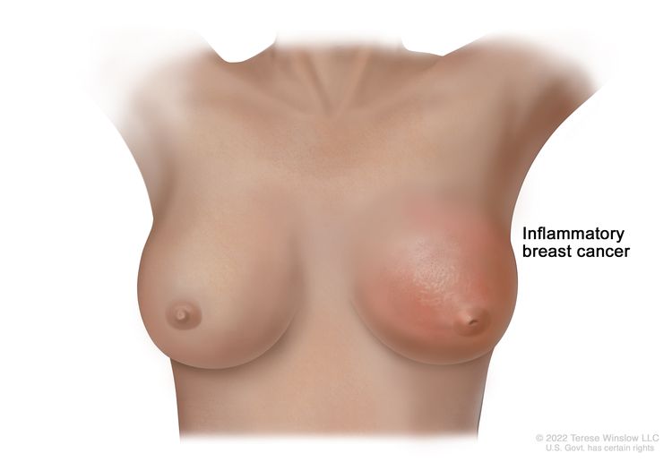
El cáncer de mama inflamatorio es un tipo de cáncer de mama en el que las células cancerosas bloquean los vasos linfáticos de la piel de la mama. Esto hace que la mama presente enrojecimiento e hinchazón. La piel también puede verse con hoyuelos o poros, como la piel de una naranja (peau d'orange), y el pezón podría estar invertido (orientado hacia dentro).
Tipos de tratamiento para el cáncer de mama
Puntos clave
- Existen diferentes tipos de tratamiento para los pacientes con cáncer de mama.
- The following types of treatment are used:
- Cirugía
- Radioterapia
- Quimioterapia
- Terapia hormonal
- Terapia dirigida
- Inmunoterapia
- New types of treatment are being tested in clinical trials.
- El tratamiento para el cáncer de mama puede producir efectos secundarios.
- Es posible que se necesiten cuidados de seguimiento.
Existen diferentes tipos de tratamiento para los pacientes con cáncer de mama.
Usted y su equipo de atención médica del cáncer trabajarán juntos para decidir su plan de tratamiento, que puede incluir más de un tipo de tratamiento. Se tendrán en cuenta muchos factores, como el estadio y el grado del cáncer, la presencia de determinados biomarcadores, su salud general y sus preferencias. El plan incluirá información sobre su cáncer, los objetivos del tratamiento, sus opciones de tratamiento y los posibles efectos secundarios, asi como la duración prevista del tratamiento.
Antes de comenzar el tratamiento, puede resultarle útil hablar con su equipo de atención médica del cáncer sobre qué esperar. Le ayudará saber qué debe hacer antes de que comience el tratamiento, cómo se sentirá durante el mismo y qué tipo de apoyo necesitará. Para obtener más información, consulte el sitio web Preguntas para el médico sobre el tratamiento.
The following types of treatment are used:
Cirugía
La mayoría de los pacientes con cáncer de mama se realizan una cirugía para extirpar el cáncer.
La biopsia del ganglio linfático centinela consiste en la extirpación del ganglio linfático centinela durante la cirugía. El ganglio linfático centinela es el primer ganglio linfático de un grupo de ganglios linfáticos que recibe el drenaje linfático del tumor primario . Es el primer ganglio linfático al que es probable que el cáncer se disemine desde el tumor primario. Se inyecta una sustancia radiactiva y/o un tinte azul cerca del tumor. La sustancia o el tinte fluye a través de los conductos linfáticos hacia los ganglios linfáticos. Se extirpa el primer ganglio linfático que recibe la sustancia o el tinte. Un patólogo examina el tejido bajo un microscopio para buscar células cancerosas. Si no se encuentran células cancerosas, puede que no sea necesario extirpar más ganglios linfáticos. A veces, se encuentra un ganglio linfático centinela en más de un grupo de ganglios. Después de la biopsia del ganglio linfático centinela, el cirujano extirpa el tumor mediante cirugía conservadora de mama o mastectomía . Si se encuentran células cancerosas, se extirparán más ganglios linfáticos a través de una incisión (corte) separada. Esto se denomina disección de ganglios linfáticos .
Los tipos de cirugía incluyen:
- La cirugía conservadora de la mama es una operación para extirpar el cáncer y parte del tejido normal que lo rodea, pero no la mama en sí. También puede quitarse parte del revestimiento de la pared torácica si el cáncer está en un área cercana. Este tipo de cirugía también puede llamarse lumpectomía, mastectomía parcial, mastectomía segmentaria, cuadrantectomía o cirugía conservadora de la mama.
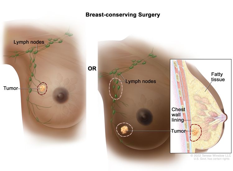
Lumpectomía. Se extirpa el tumor y parte del tejido sano circundante, pero no la mama en sí. También se pueden extirpar algunos ganglios linfáticos de la axila. Si el cáncer se encuentra cerca de la pared torácica, es posible que también se extirpe parte del revestimiento de la pared torácica.
- La mastectomía total es una cirugía para extirpar toda la mama que tiene cáncer. Este procedimiento también se llama mastectomía simple. Es posible que se extirpen algunos de los ganglios linfáticos debajo del brazo y se examinen para detectar cáncer. Esto se puede hacer al mismo tiempo que la cirugía de mama o después y se realiza a través de una incisión separada.
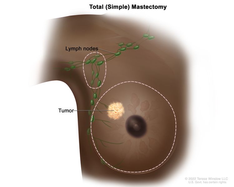
Mastectomía total (simple): se extrae toda la mama. También se pueden extirpar algunos de los ganglios linfáticos debajo del brazo.
- La mastectomía radical modificada es una cirugía para extirpar toda la mama que tiene cáncer. Esto puede incluir la extirpación del pezón, la areola (la piel de color oscuro alrededor del pezón) y la piel sobre la mama. También se extirpa la mayoría de los ganglios linfáticos debajo del brazo.
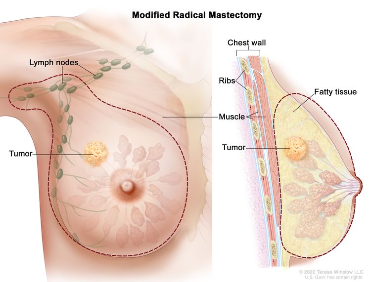
Mastectomía radical modificada: se extirpa toda la mama y la mayoría de los ganglios linfáticos debajo del brazo.
Se puede administrar quimioterapia antes de la cirugía para extirpar el tumor. Cuando se administra antes de la cirugía, la quimioterapia reducirá el tamaño del tumor y la cantidad de tejido que se debe extirpar durante la cirugía. El tratamiento administrado antes de la cirugía se llama terapia preoperatoria o terapia neoadyuvante.
Después de que el médico extirpa todo el cáncer que se puede observar en el momento de la cirugía, es posible que a algunos pacientes se les administre radioterapia, quimioterapia, terapia dirigida o terapia hormonal después de la cirugía, para destruir las células cancerosas que queden. El tratamiento administrado después de la cirugía, para reducir el riesgo de que el cáncer regrese, se llama terapia posoperatoria o terapia adyuvante.
Si una paciente se va a realizar una mastectomía, se puede considerar la reconstrucción mamaria (cirugía para reconstruir la forma de la mama después de una mastectomía). La reconstrucción mamaria puede realizarse en el momento de la mastectomía o algún tiempo después. La mama reconstruida puede fabricarse con tejido (no mamario) de la propia paciente o mediante implantes rellenos de solución salina o gel de silicona. Antes de tomar la decisión de colocarse un implante, las pacientes pueden llamar al Centro de Dispositivos y Salud Radiológica de la Administración de Alimentos y Medicamentos (FDA) al 1-888-INFO-FDA (1-888-463-6332) o visitar el sitio web de la FDA para obtener más información sobre los implantes mamarios.
Radioterapia
La radioterapia es un tratamiento contra el cáncer que utiliza rayos X de alta energía u otros tipos de radiación para matar las células cancerosas o evitar que crezcan. Hay dos tipos de radioterapia:
- La radioterapia externa utiliza una máquina que envía radiación desde el exterior del cuerpo hacia la zona donde se encuentra el cáncer.
- La radioterapia interna utiliza una sustancia radiactiva sellada en agujas, semillas, alambres o catéteres que se colocan directamente dentro o cerca del cáncer.
La forma en que se administra la radioterapia depende del tipo y estadio del cáncer que se esté tratando. La radioterapia externa se utiliza para tratar el cáncer de mama. La radioterapia interna con estroncio-89 (un radionúclido ) se utiliza para aliviar el dolor óseo causado por el cáncer de mama que se ha diseminado a los huesos. El estroncio-89 se inyecta en una vena y viaja hasta la superficie de los huesos. Se libera radiación que destruye las células cancerosas en los huesos.
Más información sobre la radiación para tratar el cáncer y los efectos secundarios de la radioterapia.
Quimioterapia
La quimioterapia (también llamada quimio) utiliza medicamentos para detener el crecimiento de las células cancerosas, ya sea destruyéndolas o impidiendo que se dividan. La quimioterapia para el cáncer de mama suele ser sistémica, es decir, se inyecta en una vena o se administra por vía oral. Cuando se administra de esta manera, los medicamentos ingresan al torrente sanguíneo para llegar a las células cancerosas en todo el cuerpo.
To learn more about how chemotherapy works, how it is given, common side effects, and more, see Chemotherapy to Treat Cancer and Chemotherapy and You: Support for People With Cancer.
Más información sobre los medicamentos aprobados para el cáncer de mama.
Terapia hormonal
La terapia hormonal (también llamada terapia endocrina) retarda o detiene el crecimiento de tumores sensibles a las hormonas al bloquear la capacidad del cuerpo para producir hormonas o al interferir con los efectos de las hormonas en las células del cáncer de mama. Las hormonas son sustancias producidas por glándulas en el cuerpo y que circulan en el torrente sanguíneo. Algunas hormonas pueden hacer que ciertos cánceres crezcan. Si las pruebas muestran que las células cancerosas tienen sitios donde las hormonas pueden adherirse (receptores), se utilizan medicamentos, cirugía o radioterapia para reducir la producción de hormonas o bloquear su acción. Esto se llama ablación ovárica.
Los tipos de terapia hormonal para el cáncer de mama son:
- terapia con inhibidores de la aromatasa (como anastrozol, letrozol o exemestano )
- Fulvestrant
- Elacestrant
- terapia con agonistas de la hormona liberadora de hormona luteinizante (LHRH) (como goserelina o leuprolida )
- Acetato de megestrol
- Tamoxifeno
Más información sobre la terapia hormonal para el cáncer de mama.
Terapia dirigida
La terapia dirigida utiliza medicamentos u otras sustancias para identificar y atacar células cancerosas específicas. Su médico puede sugerirle pruebas de biomarcadores para ayudar a predecir su respuesta a ciertos medicamentos de terapia dirigida. Más información sobre las pruebas de biomarcadores para el tratamiento del cáncer. Se utilizan varios tipos de terapia dirigida para tratar el cáncer de mama.
- los anticuerpos monoclonales son sistema inmunitario proteínas producidas en el laboratorio para tratar muchas enfermedades, entre ellas el cáncer. Como tratamiento contra el cáncer, estos anticuerpos pueden unirse a un objetivo específico en las células cancerosas u otras células que pueden ayudar a que las células cancerosas crezcan. Los anticuerpos se administran mediante infusión. Pueden usarse solos o para transportar fármacos, toxinas o material radiactivo directamente a las células cancerosas. Los anticuerpos monoclonales pueden usarse en combinación con quimioterapia como terapia adyuvante.
Los anticuerpos monoclonales utilizados para tratar el cáncer de mama son:
¿Cómo actúan los anticuerpos monoclonales para tratar el cáncer? En este video se explica cómo los anticuerpos monoclonales, como el trastuzumab, el pembrolizumab y el rituximab, bloquean moléculas que las células cancerosas necesitan para multiplicarse, marcan células cancerosas para que el sistema inmunitario las destruya o transportan sustancias que dañan estas células.
- Los inhibidores de la tirosina quinasa bloquean las señales necesarias para que crezcan los tumores. Se pueden usar en combinación con otros medicamentos contra el cáncer como la terapia adyuvante. Los inhibidores de la tirosina cinasa utilizados para tratar el cáncer de mama HER2 positivo son:
- Los inhibidores de las quinasas dependientes de ciclina (CDK) bloquean las proteínas llamadas quinasas dependientes de ciclina, que provocan el crecimiento de las células cancerosas. Los inhibidores de las CDK se pueden administrar con terapia hormonal, como Fulvestrant o Letrozol, para tratar el cáncer de mama positivo para receptores hormonales y HER2 negativo. Los inhibidores de las CDK que se utilizan para tratar el cáncer de mama son:
- Los inhibidores de la diana de rapamicina en mamíferos (mTOR) bloquean una proteína llamada mTOR, que puede impedir el crecimiento de las células cancerosas y prevenir el crecimiento de nuevos vasos sanguíneos que los tumores necesitan para crecer. Los inhibidores de mTOR utilizados para tratar el cáncer de mama HER2-negativo con receptores hormonales positivos son:
- Los inhibidores de PARP bloquean la reparación del ADN y puede producir la muerte de las células cancerosas. Los inhibidores de PARP se utilizan para tratar el cáncer de mama HER2-negativo con mutaciones en la BRCA1 o BRCA2 y son:
Obtenga más información en Terapia dirigida para tratar el cáncer.
Inmunoterapia
La inmunoterapia ayuda al sistema inmunitario de una persona a combatir el cáncer. Su médico puede sugerirle que se haga pruebas de biomarcadores para ayudar a predecir su respuesta a ciertos medicamentos de inmunoterapia. Más información sobre las pruebas de biomarcadores para el tratamiento del cáncer.
Los inhibidores de puestos de control inmunitario son un tipo de inmunoterapia que se utiliza para tratar el cáncer de mama:
- Los inhibidores de puestos de control inmunitario bloquean a las proteínas llamadas puntos de control que son producidas por algunos tipos de células del sistema inmunitario, como las células Ty algunas células cancerosas. Estos puestos de control ayudan a impedir que las respuestas inmunes sean demasiado fuertes, y a veces pueden impedir que las células T destruyan las células cancerosas. Cuando estos puestos de control se bloquean, las células T pueden destruir células cancerosas con mayor efectividad. Los inhibidores de puestos de control inmunitarios que se usan para tratar el cáncer de mama son:
Este medicamento actúa en más de una forma para matar las células cancerosas. También se considera una terapia dirigida porque se dirige a cambios o sustancias específicas en las células cancerosas.
La inmunoterapia utiliza el sistema inmunitario del cuerpo para combatir el cáncer. Esta animación explica un tipo de inmunoterapia que utiliza inhibidores de puestos de control inmunitarios para tratar el cáncer.
Más información sobre la inmunoterapia para tratar el cáncer y los efectos secundarios de la inmunoterapia.
New types of treatment are being tested in clinical trials.
For some people, joining a clinical trial may be an option. There are different types of clinical trials for people with cancer. For example, a treatment trial tests new treatments or new ways of using current treatments. Supportive care and palliative care trials look at ways to improve quality of life, especially for those who have side effects from cancer and its treatment.
You can use the clinical trial search to find NCI-supported cancer clinical trials accepting participants. The search allows you to filter trials based on the type of cancer, your age, and where the trials are being done. Clinical trials supported by other organizations can be found on the ClinicalTrials.gov website.
Para más información sobre ensayos clínicos, cómo encontrarlos y participar en uno de ellos, visite la web Información sobre estudios clínicos para pacientes y cuidadores.
El tratamiento para el cáncer de mama puede producir efectos secundarios.
Para obtener más información sobre los efectos secundarios que aparecen durante el tratamiento del cáncer, consulte la sección Efectos secundarios.
Algunos tratamientos para el cáncer de mama pueden causar efectos secundarios que continúan o aparecen meses o años después de finalizar el tratamiento. Estos se denominan efectos tardíos.
Los efectos tardíos de la radioterapia no son comunes, pero pueden ser:
- Inflamación del pulmón después de la radioterapia dirigida a la mama, especialmente cuando se administra quimioterapia al mismo tiempo.
- Linfedema del brazo, especialmente cuando se administra radioterapia después de la disección de los ganglios linfáticos. Para obtener más información, consulte Linfedema.
- En mujeres menores de 45 años que reciben radioterapia en la pared torácica después de una mastectomía, puede haber un mayor riesgo de desarrollar cáncer en la otra mama.
Los efectos tardíos de la quimioterapia dependen de los medicamentos utilizados, pero pueden ser:
Los efectos tardíos de la terapia dirigida con trastuzumab, lapatinib o pertuzumab pueden ser:
- Problemas cardiacos, como insuficiencia cardiaca
Es posible que se necesiten cuidados de seguimiento.
Algunas de las pruebas realizadas para diagnosticar o estadificar el cáncer pueden repetirse para evaluar la eficacia del tratamiento. Las decisiones sobre si continuar, modificar o suspender el tratamiento pueden basarse en los resultados de estas pruebas. A estas pruebas se las suele llamar pruebas de seguimiento o revisiones.
Tratamiento del cáncer de mama en estadio temprano, localizado u operable
For information about the treatments listed below, see the Treatment Option Overview section.
El tratamiento del cáncer de mama en etapa temprana, localizado u operable puede incluir:
Cirugía
- Cirugía conservadora de mama y biopsia de ganglio centinela. Si se encuentra cáncer en los ganglios linfáticos, se puede realizar una disección de los ganglios linfáticos.
- Mastectomía radical modificada También se puede realizar una reconstrucción mamaria.
Radioterapia posoperatoria
En el caso de las mujeres que se realizaron una cirugía de conservación del seno, se administra radioterapia en toda la mama para reducir la posibilidad de que el cáncer regrese. También se puede administrar radioterapia a los ganglios linfáticos del área.
En el caso de las mujeres que se realizaron una mastectomía radical modificada, se puede administrar radioterapia para disminuir la posibilidad de que el cáncer regrese si se cumple alguna de las siguientes condiciones:
- Se encontró cáncer en cuatro o más ganglios linfáticos.
- El cáncer se ha extendido al tejido alrededor de los ganglios linfáticos.
- El tumor era grande.
- Hay un tumor cerca o que permanece en el tejido cerca de los bordes de donde se extirpó el tumor.
Terapia sistémica posoperatoria
La terapia sistémica es el uso de medicamentos que pueden ingresar al torrente sanguíneo y llegar a las células cancerosas de todo el cuerpo. La terapia sistémica posoperatoria se administra para disminuir la posibilidad de que el cáncer regrese después de la cirugía para extirpar el tumor.
La terapia sistémica posoperatoria se administra en función de si:
- El tumor es negativo o positivo para receptores hormonales.
- El tumor es HER2 negativo o positivo.
- El tumor es negativo para receptores hormonales y negativo para HER2 ( triple negativo ).
- El tamaño del tumor
En mujeres premenopáusicas con tumores con receptores hormonales positivos, es posible que no se necesite más tratamiento, o que la terapia postoperatoria incluya:
- Terapia con tamoxifeno con o sin quimioterapia
- El tratamiento con tamoxifeno busca detener o disminuir la producción de estrógeno en los ovarios . También se puede recurrir a la terapia farmacológica, la cirugía para extirpar los ovarios o la radioterapia ovárica.
- Terapia y tratamiento con inhibidores de la aromatasa para detener o disminuir la cantidad de estrógeno que producen los ovarios. Se puede utilizar terapia con medicamentos, cirugía para extirpar los ovarios o radioterapia en los ovarios.
En mujeres posmenopáusicas con tumores con receptores hormonales positivos, es posible que no se necesite más tratamiento, o que la terapia postoperatoria incluya:
- Terapia con inhibidores de la aromatasa con o sin quimioterapia
- Tamoxifeno seguido de terapia con inhibidores de la aromatasa, con o sin quimioterapia
En mujeres con tumores con receptores hormonales negativos, es posible que no se necesite más tratamiento, o que la terapia postoperatoria incluya quimioterapia.
En mujeres con tumores HER2 negativos, el tratamiento postoperatorio puede incluir quimioterapia.
En mujeres con tumores pequeños, HER2 positivos y sin cáncer en los ganglios linfáticos, es posible que no se necesite más tratamiento. Si hay cáncer en los ganglios linfáticos o el tumor es grande, la terapia posoperatoria puede consistir en:
- Quimioterapia y terapia dirigida (trastuzumab)
- Terapia hormonal, como tamoxifeno o terapia con inhibidores de la aromatasa, para tumores que también tienen receptores hormonales positivos.
En mujeres con tumores pequeños, negativos para receptores hormonales y HER2 (triple negativos) y sin cáncer en los ganglios linfáticos, es posible que no se necesite más tratamiento. Si hay cáncer en los ganglios linfáticos o el tumor es grande, la terapia postoperatoria puede incluir:
- Chemotherapy.
- Radioterapia
- Terapia con inhibidores de PARP para mujeres con una mutación hereditaria en los genes BRCA1 o BRCA2 .
- Un ensayo clínico de un nuevo régimen de quimioterapia
Terapia sistémica preoperatoria
La terapia sistémica consiste en el uso de medicamentos que pueden ingresar al torrente sanguíneo y llegar a las células cancerosas en todo el cuerpo. La terapia sistémica preoperatoria se administra para reducir el tamaño del tumor antes de la cirugía.
La quimioterapia preoperatoria puede hacer posible la cirugía para conservar la mama en pacientes que de otro modo no serían elegibles. La quimioterapia preoperatoria también puede reducir la necesidad de realizar una disección de ganglios linfáticos en pacientes con enfermedad que se ha diseminado a los ganglios linfáticos.
En mujeres posmenopáusicas con tumores con receptores hormonales positivos, la terapia preoperatoria puede consistir en:
- Chemotherapy.
- Terapia hormonal, como tamoxifeno o terapia con inhibidores de la aromatasa, para mujeres que no pueden recibir quimioterapia.
En mujeres premenopáusicas con tumores con receptores hormonales positivos, el tratamiento preoperatorio puede incluir un ensayo clínico de terapia hormonal, como tamoxifeno o terapia con inhibidores de la aromatasa.
Quimioterapia y terapia dirigida (trastuzumab)
- Quimioterapia y terapia dirigida (trastuzumab)
- Terapia dirigida (pertuzumab)
En mujeres con tumores HER2 negativos o tumores triple negativos, el tratamiento preoperatorio puede incluir quimioterapia.
Para pacientes con enfermedad triple negativa o HER2 positiva, la respuesta a la terapia preoperatoria puede usarse como guía para elegir el mejor tratamiento después de la cirugía.
Puede utilizar la búsqueda de ensayos clínicos y encontrar ensayos clínicos sobre cáncer patrocinados por el NCI que acepten participantes. La búsqueda le permite filtrar los ensayos según el tipo de cáncer, la edad y el lugar donde se realizan los ensayos. También encontrará información general sobre los ensayos clínicos.
Tratamiento del cáncer de mama localmente avanzado o inflamatorio
For information about the treatments listed below, see the Treatment Option Overview section.
El tratamiento del cáncer de mama localmente avanzado o inflamatorio es una combinación de terapias que pueden incluir:
- Cirugía ( cirugía conservadora de mama o mastectomía total ) con disección de ganglios linfáticos .
- Quimioterapia antes y/o después de la cirugía
- Radioterapia después de la cirugía
- Terapia hormonal después de la cirugía para tumores con receptores de estrógeno positivos o con receptores de estrógeno desconocidos.
- Terapia dirigida (trastuzumab y pertuzumab)
- Ensayos clínicos que evalúan nuevos medicamentos contra el cáncer, nuevas combinaciones de medicamentos y nuevas formas de administrar tratamientos.
Puede utilizar la búsqueda de ensayos clínicos y encontrar ensayos clínicos sobre cáncer patrocinados por el NCI que acepten participantes. La búsqueda le permite filtrar los ensayos según el tipo de cáncer, la edad y el lugar donde se realizan los ensayos. También encontrará información general sobre los ensayos clínicos.
Tratamiento del cáncer de mama recidivante locorregional
For information about the treatments listed below, see the Treatment Option Overview section.
El tratamiento del cáncer de mama recurrente locorregional (cáncer que ha reaparecido después del tratamiento en la mama, en la pared torácica o en los ganglios linfáticos cercanos) puede incluir:
- Quimioterapia.
- Terapia hormonal para tumores con receptores hormonales positivos .
- Radioterapia.
- Cirugía
- Terapia dirigida (trastuzumab y pertuzumab)
- Participación en el ensayo clínico de un tratamiento nuevo
Para obtener información sobre las opciones de tratamiento para el cáncer de mama que se ha propagado a partes del cuerpo fuera de la mama, la pared torácica o los ganglios linfáticos cercanos, consulte la sección Tratamiento del cáncer de mama metastásico.
Puede utilizar la búsqueda de ensayos clínicos y encontrar ensayos clínicos sobre cáncer patrocinados por el NCI que acepten participantes. La búsqueda le permite filtrar los ensayos según el tipo de cáncer, la edad y el lugar donde se realizan los ensayos. También encontrará información general sobre los ensayos clínicos.
Tratamiento del cáncer de mama metastásico
For information about the treatments listed below, see the Treatment Option Overview section.
Las opciones de tratamiento para el cáncer de mama metastásico (cáncer que se ha diseminado a partes distantes del cuerpo) pueden incluir:
Terapia hormonal
En mujeres posmenopáusicas a las que se les acaba de diagnosticar cáncer de mama metastásico con receptores hormonales positivos o si se desconoce el estado de los receptores hormonales, el tratamiento puede incluir:
- Terapia con tamoxifeno .
- Terapia con inhibidores de la aromatasa (anastrozol, letrozol o exemestano). A veces también se administra terapia con inhibidores de la quinasa dependiente de ciclina (palbociclib, ribociclib, abemaciclib o alpelisib).
- Terapia con inhibidores de PARP para mujeres con una mutación hereditaria en los genes BRCA1 o BRCA2 .
En mujeres premenopáusicas a las que se les acaba de diagnosticar cáncer de mama metastásico positivo para receptores hormonales, el tratamiento puede consistir en:
- Tamoxifeno, un agonista de la LHRH o ambos
- Tratamiento con inhibidores de las quinasas dependientes de ciclinas (ribociclib)
En mujeres cuyos tumores son receptores hormonales positivos o receptores hormonales desconocidos, con metástasis solo en el hueso o los tejidos blandos, y que han sido tratadas con tamoxifeno, el tratamiento puede incluir:
- Terapia con inhibidores de la aromatasa
- Otras terapias hormonales como el acetato de megestrol, la terapia con estrógenos o andrógenos, o la terapia antiestrogénica como el fulvestrant o el elacestrant .
Terapia dirigida
En mujeres con cáncer de mama metastásico positivo para receptores hormonales que no ha respondido a otros tratamientos, las opciones pueden consistir en terapia dirigida como:
- Trastuzumab, lapatinib, pertuzumab o inhibidores de mTOR
- Terapia con inhibidores de la cinasa dependiente de ciclina (palbociclib, ribociclib o abemaciclib), que puede combinarse con terapia hormonal.
En mujeres con cáncer de mama metastásico HER2 positivo, el tratamiento puede consistir en:
- Terapia dirigida como trastuzumab, trastuzumab deruxtecan, pertuzumab, margetuximab o lapatinib
- Terapia dirigida con tucatinib, un inhibidor de la tirosina quinasa que se usa con trastuzumab y capecitabina
En mujeres con cáncer de mama metastásico HER2 negativo, con mutaciones en los genes BRCA1 o BRCA2, y que han sido tratadas con quimioterapia, el tratamiento puede consistir en terapia dirigida con un inhibidor de PARP (olaparib o talazoparib).
Quimioterapia
En las mujeres con cáncer de mama metastásico que no ha respondido a la terapia hormonal, se ha diseminado a otros órganos o ha causado síntomas, el tratamiento puede consistir en quimioterapia con uno o más medicamentos.
Quimioterapia e inmunoterapia
En mujeres con tumores de mama triple negativos localmente recidivantes, inoperables o metastásicos que expresan PD-L1, el tratamiento puede consistir en quimioterapia e inmunoterapia (pembrolizumab).
Cirugía
- Mastectomía total para mujeres con lesiones mamarias abiertas o dolorosas. Se puede administrar radioterapia después de la cirugía.
- Cirugía para extirpar el cáncer que se ha diseminado al cerebro o la columna vertebral. Se puede administrar radioterapia después de la cirugía.
- Cirugía para extirpar el cáncer que se ha diseminado al pulmón.
- Cirugía para reparar o reforzar huesos débiles o fracturados. La radioterapia se puede administrar después de la cirugía.
- Cirugía para eliminar el líquido que se ha acumulado alrededor de los pulmones o el corazón.
Radioterapia
- Radioterapia dirigida a los huesos, el cerebro, la médula espinal, la mama o la pared torácica para aliviar los síntomas y mejorar la calidad de vida.
- Estroncio-89 (un radionúclido) para aliviar el dolor producido por el cáncer que se ha diseminado a los huesos de todo el cuerpo.
Otras opciones de tratamiento
Otras opciones de tratamiento para el cáncer de mama metastásico pueden ser:
- Terapia farmacológica con bifosfonatos o denosumab para mitigar la enfermedad ósea y el dolor cuando el cáncer se ha propagado a los huesos. Para obtener información sobre los bifosfonatos, consulte la página web de dolor por cáncer.
- Terapia conjugada anticuerpo-fármaco con sacituzumab govitecan para ciertas pacientes con cáncer de mama triple negativo metastásico. Sacituzumab govitecan también está aprobado para algunas pacientes con cáncer de mama metastásico con receptores hormonales positivos y HER2 negativo.
- Un ensayo clínico de quimioterapia en dosis altas con trasplante de células madre
- Ensayos clínicos que evalúan nuevos medicamentos para el cáncer, nuevas combinaciones de medicamentos y nuevas formas de administrar tratamientos.
Puede utilizar la búsqueda de ensayos clínicos y encontrar ensayos clínicos sobre cáncer patrocinados por el NCI que acepten participantes. La búsqueda le permite filtrar los ensayos según el tipo de cáncer, la edad y el lugar donde se realizan los ensayos. También encontrará información general sobre los ensayos clínicos.
Tratamiento del carcinoma ductal in situ (CDIS)
For information about the treatments listed below, see the Treatment Option Overview section.
El tratamiento del carcinoma ductal in situ puede consistir en:
- Cirugía conservadora de mama y radioterapia, con o sin tamoxifeno
- Mastectomía total con o sin tamoxifeno. También se puede administrar radioterapia.
Puede utilizar la búsqueda de ensayos clínicos y encontrar ensayos clínicos sobre cáncer patrocinados por el NCI que acepten participantes. La búsqueda le permite filtrar los ensayos según el tipo de cáncer, la edad y el lugar donde se realizan los ensayos. También encontrará información general sobre los ensayos clínicos.
Más información sobre el cáncer de mama
Para obtener más información del National Cancer Institute sobre el cáncer de mama, consulte las siguientes páginas web:
- Página de inicio sobre el cáncer de mama
- Opciones de cirugía para mujeres con CDIS o cáncer de mama
- Cirugía para reducir el riesgo de desarrollar cáncer de mama
- Reconstrucción mamaria después de la mastectomía
- Biopsia de ganglio linfático centinela
- Mamas densas: respuestas a las preguntas más frecuentes
- Medicamentos aprobados para el cáncer de mama
- Terapia hormonal para el cáncer de mama
- Terapia dirigida para tratar el cáncer
- Immunotherapy to Treat Cancer
- Cáncer de mama inflamatorio
- Alteraciones en el gen BRCA: riesgo de cáncer y pruebas genéticas
- Pruebas genéticas para síndromes hereditarios de susceptibilidad al cáncer
For general cancer information and other resources from the National Cancer Institute, visit:
Sobre este resumen del PDQ
Acerca del PDQ
El Physician Data Query (PDQ) es la base de datos integral sobre el cáncer del National Cancer Institute (NCI). La base de datos del PDQ contiene resúmenes con la última información publicada sobre prevención, detección, genética, tratamiento, atención médica de apoyo y medicina complementaria y alternativa relacionada con el cáncer. La mayoría de los resúmenes se presentan en dos versiones. Las versiones para profesionales de la salud contienen información detallada escrita en lenguaje técnico. Las versiones para pacientes están escritas en un lenguaje fácil de entender y no tan técnico. Ambas versiones contienen información precisa y actualizada sobre el cáncer. La mayoría de las versiones también están disponibles en español.
El PDQ es un servicio del NCI. El NCI es parte de los Institutos Nacionales de Salud (NIH), que son el centro de investigación biomédica del Gobierno federal. Los resúmenes del PDQ se basan en una revisión independiente de la literatura médica. No son declaraciones de políticas del NCI ni de los NIH.
Propósito de este resumen
Este resumen del PDQ sobre el cáncer contiene información actualizada sobre el tratamiento del cáncer de mama en adultos. Su propósito es informar y ayudar a los pacientes, las familias y los cuidadores. No da pautas ni recomendaciones formales para tomar decisiones relacionadas con la atención médica.
Revisores y actualizaciones
Los comités editoriales escriben los resúmenes de información sobre el cáncer del PDQ y los mantienen actualizados. Estos comités están formados por equipos de especialistas en el tratamiento del cáncer y otras especialidades relacionadas con esta enfermedad. Los resúmenes se revisan periódicamente y se modifican cuando hay información nueva. La fecha de actualización al pie de cada resumen indica cuándo se realizó el cambio más reciente.
The information in this patient summary was taken from the health professional version, which is reviewed regularly and updated as needed, by the PDQ Adult Treatment Editorial Board.
Información sobre ensayos clínicos
Un ensayo clínico es un estudio para responder a una pregunta científica como, por ejemplo, si un tratamiento es mejor que otro. Los ensayos se basan en estudios anteriores y en lo aprendido en el laboratorio. Cada ensayo responde a determinadas preguntas científicas que permiten encontrar nuevas y mejores formas de ayudar a los pacientes con cáncer. Durante los ensayos clínicos de tratamiento, se recopila información sobre los efectos de un nuevo tratamiento y su eficacia. Si un ensayo clínico demuestra que un nuevo tratamiento es mejor que uno que se utiliza actualmente, el nuevo tratamiento puede convertirse en “estándar”. Los pacientes pueden valorar la posibilidad de participar en un ensayo clínico. Algunos ensayos clínicos solo están abiertos a pacientes que no hayan iniciado el tratamiento.
Los ensayos clínicos se pueden encontrar en línea en el sitio web del NCI. Para obtener más información, llame al Servicio de Información sobre el Cáncer (CIS, por sus siglas en inglés), el centro de contacto del NCI, al 1-800-4-CANCER (1-800-422-6237).
Permiso de uso de este resumen
Physician Data Query (PDQ) es una marca registrada. Se autoriza el libre uso del contenido de los documentos del PDQ como texto. Sin embargo, no se podrá identificar como un resumen de información sobre cáncer del PDQ del NCI, salvo que se reproduzca en su totalidad y se actualice con regularidad. Por otra parte, se permite que los autores incluyan una oración como “en el resumen del PDQ del NCI sobre la prevención del cáncer de mama se describen, de manera concisa, los siguientes riesgos: [incluir fragmento del resumen]”.
La forma recomendada para citar este resumen del PDQ es:
Comité editorial del PDQ® sobre el tratamiento para adultos. Tratamiento del cáncer de mama (PDQ). Bethesda, MD: National Cancer Institute. Actualizado el [DD/MM/AAAA]
Las imágenes de este resumen se utilizan con el permiso del autor, artista y/o editorial para uso exclusivo en los resúmenes del PDQ. Si desea usar una imagen de un resumen del PDQ sin incluir el resumen completo, debe obtener autorización del propietario. El National Cancer Institute no puede otorgar dicho permiso. Para obtener más información sobre el uso de las imágenes de este resumen o de otras ilustraciones relacionadas con el cáncer, consulte Visuals Online, una colección de más de 3,000 imágenes científicas.
Descargo de responsabilidad
La información de estos resúmenes no debe utilizarse para tomar decisiones sobre reembolsos de seguros. Puede encontrar más información sobre la cobertura de seguros en Cancer.gov en el sitio Manejo de la atención del cáncer.
Contáctenos
Puede encontrar más información sobre cómo contactarnos o recibir ayuda en el sitio web Cancer.gov en la página Comuníquese con el NCI. También puede enviar sus preguntas a Cancer.gov en el apartado Escríbanos del sitio web.
Actualizado:
URL de origen: https://www.cancer.gov/node/4127/syndication
Agencia de origen: National Cancer Institute (NCI)
Fecha de captura: 2013-09-14 09:02:02.0