


Our approach to Cardiovascular Imaging
Video could not be played
Ranked in the top 1% of all hospitals in the nation for Cardiology, Heart & Vascular Surgery according to U.S. News & World Report, our world-renowned specialists are passionate about uncovering the latest diagnostic approaches and treatments that can improve outcomes.
Cardiovascular imaging is an essential component of cardiovascular diagnostics. The Montefiore Einstein Cardiovascular Imaging Program is among the largest in New York City, with 60,000 imaging studies performed each year, using the most advanced technology available. Our program works in a multidisciplinary team to create baseline imaging heart studies. This allows our cardiologists and our cardiothoracic and vascular surgeons to offer a comprehensive diagnosis and individualized treatment plans for all heart conditions.
Working together with our renowned cardiologists and cardiothoracic surgeons, we provide the most precise diagnoses possible using non-invasive methods, advanced technology in cardiovascular intervention, and state-of-the-art resources. We are committed to achieving the best clinical outcomes for outpatient therapies, non-invasive interventions and surgery alike.
Video could not be played
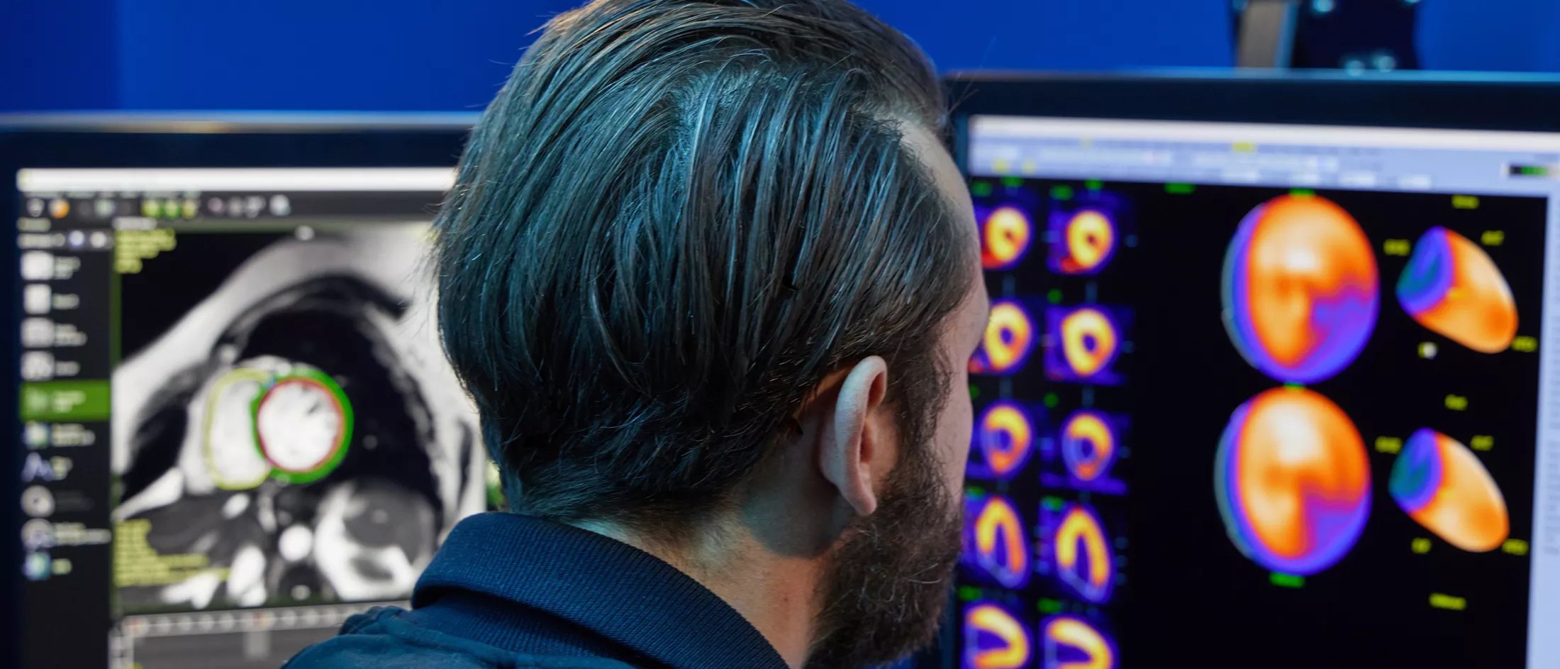
Advanced Imaging
We offer cardiovascular imaging for the full spectrum of cardiac and cardiovascular conditions, utilizing the most advanced techniques to achieve the best results and most thorough diagnoses.
A noninvasive test that provides cardiologists with a survey of the heart with the same ultrasound technology used for pregnant women. The echocardiograph, or echo, allows cardiologists to examine the size and function of a patient's heart and the condition of the patient's valves to determine how efficiently the heart contracts. Echos also reveal common congenital problems and possible causes of murmurs, palpitations, chest pain and shortness of breath. During the 20-30 minute exam, the sonographer will take still pictures of the heart, record video of the heart beating, and examine blood flow.
The stress echo gives the cardiologist a picture of the heart after the patient has been exercising on a treadmill. By comparing a stress echo to an at-rest echo, the cardiologist may catch issues that surface only when the heart is under stress. Roughly 95% of stress echos are performed to look for evidence of blocked arteries in patients experiencing chest pain, shortness of breath or palpitations. For patients who are unable to walk on the treadmill due to disability or lung conditions, the pharmacological stress echo can be administered with Dobutamine, a medication that increases the heart muscle’s need for oxygen as well as the heart pump function without exercise.
When a patient is unable to exercise or take dobutamine, the nuclear stress test can be administered with adenosine instead of dobutamine. The procedure is longer than a traditional stress echo test, and the results are not available as quickly because they require processing by technicians and further assessment by our cardiologists.
Unlike an echo, which is non-invasive, transesophageal echocardiography (TEE) is performed by inserting a probe into the esophagus. This view through the esophagus offers a clearer picture of the heart than a traditional surface echo. Transesophageal echocardiography can be performed in cardiac catheterization laboratories and operating rooms, and they assist cardiac interventionists and surgeons to better treat your heart disease.
Advanced Imaging: A Non-invasive Alternative
Our cardiology team continuously seeks the best, newest techniques and implements the most advanced technology for imaging the heart, including: live, three-dimensional echocardiography, myocardial strain, high-resolution cardiac computed tomography and cardiac magnetic resonance to diagnose structural heart disease, coronary artery disease and specific cardiomyopathy, such as amyloidosis, sarcoidosis, low pump function condition. We are currently developing 3D prints to better understand structural heart disease.
Computed tomography (CT) is the first test to give doctors a truly three-dimensional picture of how a patient's coronary arteries run along the surface of the heart. This test is the latest advance in the effort to map blockages in coronary arteries and congenital abnormalities. In general, when a block is suspected, cardiologists recommend that patients undergo an angiogram, an invasive test that probes the coronary arteries. CT angiography, however, gives cardiologists a clear picture of the actual block, which may eventually lead to a reduction in the number of patients requiring invasive testing.
Cardiac magnetic resonance imaging (MRI) is used to search for tearing and aneurysm or bulging of the aorta. MRI is an effective tool for mapping the overall heart structure of patients with congenital heart problems. For patients who are potential candidates for coronary artery bypass surgery or for angioplasty, MRI can guide cardiologists to healthy heart tissue, which helps determine if overall function will improve with procedures to increase blood flow to the heart.
Many structure-related heart diseases were traditionally retreated with open heart surgeries which often have higher mortality and morbidity as well as longer hospital stays. The expert team at Montefiore Einstein Heart and Vascular Care Center is able to image heart valves for non-surgical intervention. Our world-renowned physicians and staff possess an in-depth knowledge and commitment to the field which allows us to provide the best possible patient care for even the most challenging conditions.
Our imaging team has focused on early identification of heart function in cancer patients who receive chemotherapy. Through early detection of occult heart function using advanced cardiovascular imaging, medical therapy can be initiated earlier to reduce cardiovascular complications.
Although present in many people, cardiac amyloidosis has historically been difficult to diagnose. Symptoms of amyloidosis mimic different diseases and the average diagnosis could take as long as 18-24 months. Our cardiovascular imaging team is developing an imaging program for amyloidosis. We use echocardiography myocardial strain imaging for early identification, followed by cardiac MRI and nuclear imaging for further classification.

Video could not be played
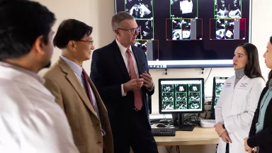
Your Cardiovascular Imaging Team

Video could not be played