


Our Approach to Thoracic Surgery
Video could not be played
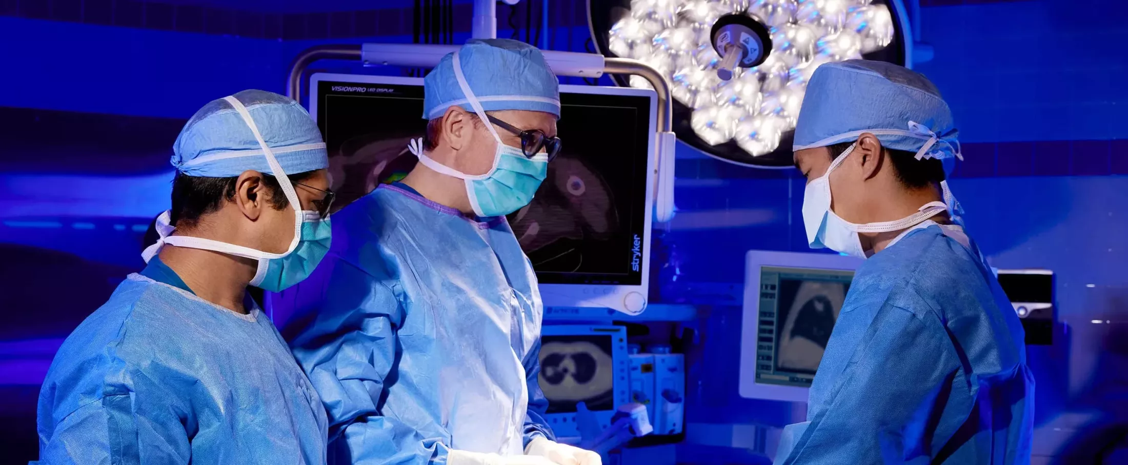
The world-renowned thoracic surgeons at Montefiore Einstein have expertise in treatment for diseases of the lung, esophagus and mediastinum. To provide world-class thoracic surgical oncology and general thoracic surgery, we focus on state-of-the-art surgical care, early detection, multidisciplinary care, and treatment plans that are fully personalized for our patients. Our new bronchoscopy suite is fully equipped with the most advanced diagnostic and therapeutic technologies, which will have a major impact on screening for lung cancer and other lung diseases.
Clinical Excellence
We are dedicated to increasing the quality of care and improving the accessibility of surgical care. Our program also fosters initiatives to increase the early detection of cancers and improve the delivery of multidisciplinary care. An integral part of excellence in care is our partnership with the exceptional cardiac surgery team at Montefiore Einstein Center for Heart and Vascular Care. For patients with cancer, we work as a team with our Comprehensive Cancer Center, which is among the elite 1%, NCI-designated comprehensive cancer centers in the U.S.
Video could not be played
State-of-the-Art Diagnostics
To develop a personalized course of treatment, our multidisciplinary team uses a range of technologies, including state-of-the-art imaging, minimally invasive biopsy and other diagnostics. Our High Risk Lung Nodule Clinic and Incidental Pulmonary Nodule Program use an automated, AI-based program to identify and track patients found to have incidental lung nodules.
In a biopsy, a sample of tissue is removed so that it can be examined under a microscope for signs of disease. A lung biopsy removes a small piece of lung tissue. A lymph node biopsy removes lymph node tissue. A pleural biopsy removes a portion of the thin tissue that lines the lungs and chest cavity. A biopsy can be performed with a bronchoscope, a needle, open surgery or video-assisted thoracoscopic surgery (VATS). The method used depends on where the sample will be taken from and the patient’s overall health.
This procedure is used to examine the breathing passages of the lungs. Flexible bronchoscopy, also called an airway endoscopy, uses a thin, tube-like instrument with a camera that is passed through the nose or mouth to reach the breathing passages of the lungs. Rigid bronchoscopy uses a straight, hollow metal tube, and it can be used to perform procedures in the airways. Montefiore Einstein’s new bronchoscopy suite is fully equipped with the most advanced diagnostic and therapeutic technologies, which will have a major impact on screening for lung cancer and other lung diseases.
Ultrasound is a common test that uses sound waves to create pictures of your internal organs and tissues. Endobronchial ultrasound creates images of your lungs and nearby lymph nodes by using an ultrasound probe housed in a flexible tube that goes through the mouth and into the windpipe and lungs. The resulting images are used to help identify areas in which to perform biopsies.
An EGD is performed using an endoscope (a flexible tube with a camera) to look inside your stomach and the first section of your intestine for abnormalities. An EGD may be used to find the cause of unexplained symptoms, identify diseases, remove polyps or other growths and treat bleeding from ulcers or other conditions. You may be given a sedative or other medication to help you stay comfortable during the procedure. During the EGD, the doctor may use the endoscope to take tissue, fluid or cell samples for testing, or perform other procedures.
Interventional esophagoscopy is used to diagnose and treat disorders of the esophagus, the passage from the throat to the stomach. This procedure uses an endoscope or esophagoscope (a thin, tube-like instrument with a light and a lens for viewing). The scope is inserted through the mouth or nose and down the throat into the esophagus, so the doctor may look for abnormal areas. The scope may also be equipped with a tool to remove tissue samples to be examined under a microscope for signs of cancer.
This surgical procedure allows the doctor to view the organs inside the abdomen for signs of disease by using a laparoscope (a thin, tube-like instrument with a light and camera). Small incisions are made in the wall of the abdomen, and the laparoscope is inserted into one of the incisions. The doctor can also use other instruments inserted through the same or other incisions to perform procedures such as taking tissue samples to be checked under a microscope for signs of disease.
This procedure is used to examine the mediastinum, the space behind the breastbone and between the lungs. A small incision is made on the chest or neck, allowing the doctor to insert a flexible tube with a camera to view the area. This procedure is used to look for signs of infection, inflammation or cancer. This procedure can also be used to perform biopsies of lymph nodes around the airway for the purpose of staging lung cancer.
A thoracoscopy enables doctors to view the organs inside the chest to check for abnormalities. It may also be used to remove tissue or lymph node samples or for surgical treatment. Small incisions are made between the ribs and a thoracoscope (a thin, tube-like instrument with a light and a camera) is inserted through an incision, providing the surgeon with a clear, magnified view of the area. Video-assisted thoracoscopic surgery (VATS) uses the thoracoscope as part of minimally invasive surgery. Robotic-assisted thoracoscopy uses a robotic surgical system that includes a thoracoscope and multiple tiny instruments, controlled in real-time by the surgeon.
Advanced Treatments
The world-renowned thoracic surgeons at Montefiore Einstein have expertise in treatment for diseases of the lung, esophagus and mediastinum. By using minimally invasive lung surgery when appropriate, including video-assisted thoracoscopic surgery (VATS) and robotic-assisted surgery, we are able to preserve vital lung tissue, lower risks of complications, and speed recovery.
When a lung nodule is detected, acting quickly is key to achieving the best outcome. That’s why the Montefiore Einstein Comprehensive Cancer Center created the Follow-Up ASessmenT of Lung Nodules (FAST) Clinic—to swiftly find, diagnose, and treat lung nodules. Equipped with a state-of-the-art robotic bronchoscopy suite, the center is able to assess and determine next steps with unmatched speed. If a diagnosis of lung or chest cancer is made, our specialists will provide a recommended treatment plan personalized for you.
Bronchoscopy/Endoscopy
Interventional bronchoscopy uses a bronchoscope (a thin, tube-like instrument with a light, a lens for viewing and other instruments, as needed) to enable minimally invasive treatment for lung tumors and other respiratory conditions, such as severe asthma and emphysema. The bronchoscope is inserted through the nose or mouth into the trachea and lungs, providing the surgeon with access to treat the affected area. Montefiore Einstein’s new bronchoscopy suite is fully equipped with the most advanced diagnostic and therapeutic technologies, which will have a major impact on screening for lung cancer and other lung diseases.
Interventional esophagoscopy is used to diagnose and treat disorders of the esophagus, the passage from the throat to the stomach This procedure uses an endoscope or esophagoscope (a thin, tube-like instrument with a light and a lens for viewing). The scope is inserted through the mouth or nose and down the throat into the esophagus, so the doctor may look for abnormalities. The scope may also be equipped with a tool to remove tissue samples to be examined under a microscope for signs of cancer.
Esophageal & Upper GI
The esophagus, which connects your throat and your stomach, can sometimes develop an abnormal outpouching or pocket, called an esophageal diverticulum. Esophageal diverticulectomy is a surgical procedure where this outpouching of the esophagus is removed to improve swallowing.
In people with achalasia, a rare swallowing disorder, the muscles of the esophagus (the tube connecting the throat and the stomach) do not contract properly and the ring of muscle at the bottom of the esophagus does not relax to let the food into the stomach. Esophageal myotomy is a surgical procedure where the affected muscle at the bottom of the esophagus is cut to enable food to flow more easily into the stomach. This treatment can often be performed through minimally invasive surgery.
An esophagectomy is a surgical procedure to remove a portion of the esophagus (the tube that connects the throat and the stomach). To enable swallowing, the doctor will connect the remaining healthy section of the esophagus to the stomach, using a plastic tube or part of the intestine, if necessary. This procedure is often used to treat cancers of the esophagus or other disorders.
Hiatal hernia occurs when part of the stomach bulges through the wall of muscle that separates the abdominal cavity from the chest cavity. Paraesophageal hernia is a type of hiatal hernia where much of the stomach protrudes into the chest cavity. While those with mild to moderate symptoms may not need treatment, severe cases may need surgery to address the condition.
Mediastinum
In this surgical procedure, sympathetic nerves are cut or clamped to treat excessive sweating, chronic pain, irregular heart rhythms or other problems with the sympathetic nervous system. This can often be completed as a minimally invasive procedure using small incisions and performed on an outpatient basis.
Operative Techniques
This robotic system provides doctors with a clear, magnified view of the organs inside the chest and enables them to conduct precise surgery, including the removal of tissue or lymph nodes. It is minimally invasive, relying on small incisions (cuts) between the ribs to insert multiple tiny instruments, including a thoracoscope (a thin, tube-like instrument with a light and a camera). With a magnified view and versatile instruments, the surgeon is able to make precise, highly controlled movements.
A thoracotomy is performed through an incision (cut) between the ribs, enabling the surgeon to see and reach the lungs or other organs in the thoracic area. The procedure is performed under general anesthesia, with a breathing tube placed in the patient’s airway, and the patient positioned on their side. Patients can expect some pain when taking a deep breath after the procedure and will be prescribed pain medication and breathing exercises during recovery. This surgery is performed to diagnose or treat a disease, and it enables the surgeon to look at, biopsy and remove tissue.
This state-of-the-art, minimally invasive surgery enables doctors to view the organs inside the chest and to remove tissue or lymph node samples. Small incisions (cuts) are made between the ribs, and a thoracoscope (a thin, tube-like instrument with a light and a camera) is inserted into the chest, providing a clear, magnified view.
Pleura & Chest Wall
This is a surgical procedure to remove a damaged or diseased portion of the chest wall.The procedure is performed under general anesthesia, with a breathing tube placed in the patient’s airway. The protective structure of the chest wall may be reconstructed using artificial bone or titanium rib plating. This surgery is most commonly performed to address cancer, infections and trauma.
This is a surgical procedure to remove part or all of the pleura, a thin membrane of tissue that lines the lungs and chest cavity. This procedure is typically performed to treat mesothelioma, a cancer that forms in the thin tissue that lines many internal organs, or other conditions such as a fluid buildup between the layers of tissue that line the lungs and chest cavity.
This procedure is used to place medicine within the chest cavity to reduce or prevent fluid buildup. The medicine triggers an inflammatory reaction on the surface of the lung and inside the chest cavity, causing the surface of the lung to stick to the surface of the chest cavity and prevent fluid buildup.
This is a surgical procedure to remove abnormal fibrous tissue, which may have formed due to a buildup of excess fluid covering the lung, chest wall or diaphragm. Depending on the medical condition, the surgery can be minimally invasive or open. The procedure is performed under general anesthesia, with a breathing tube placed in the patient’s airway.
Tunneled pleural catheters (IPC) may be placed in patients who experience buildup of fluid around the lungs (malignant pleural effusions) associated with cancer. Tunneled pleural catheters are soft, silicone tubes that allow people to better manage the shortness of breath caused by recurrent malignant pleural effusions.
Pulmonary
Each lung is divided into sections called lobes: the right lung has three lobes and the left lung has two. A bilobectomy is a surgical procedure to remove two lobes of the lung. It is commonly used in the treatment of lung cancer and is only performed for tumors of the right lung, where the tumor affects two adjacent lobes. The procedure is performed under general anesthesia and may involve minimally invasive surgery or open surgery, depending on the condition.
Each lung is divided into sections called lobes: the right lung has three lobes and the left lung has two. A lobectomy is a surgical procedure to remove an affected lobe of the lung. The procedure is performed under general anesthesia and may involve minimally invasive surgery or open surgery, depending on the condition. The surgery may be used to treat cancer, infection, COPD or benign tumors.
This surgical procedure removes lymph nodes in the area between the lung and chest in the course of treating lung cancer. The procedure is performed under general anesthesia and may involve minimally invasive surgery or open surgery, depending on the condition. The number of lymph nodes removed will vary based on the individual patient's condition.
Pneumonectomy is a surgical procedure to remove the entire lung. The procedure is performed under general anesthesia as open surgery. The surgery may be used to treat a range of conditions, including infection, bronchiectasis, benign tumors, or cancer when it cannot be removed by a lobectomy.
This surgical procedure removes a significant portion of the lung—more tissue than a wedge resection and less tissue than a lobectomy—saving the tissue that is unaffected by disease. The procedure is performed under general anesthesia and may involve minimally invasive surgery or open surgery, depending on the condition. The surgery is commonly used in the treatment of lung cancer, as well as to remove benign masses found in the lungs.
This surgical procedure is used in the treatment of lung cancer that affects both one lobe of the lung and the main bronchus to that lung. The cancerous lobe and a portion of the main bronchus are removed. The remaining end of the main bronchus is rejoined with the bronchus to unaffected lobes. This surgery avoids the need for a pneumonectomy.
This surgical procedure removes a damaged or diseased portion of the trachea, the windpipe or tube that connects the voicebox to the lungs. The procedure is performed under general anesthesia and may involve minimally invasive surgery or open surgery, depending on the condition. This surgery may be used to treat benign or cancerous tumors, injury, or narrowing of the trachea.
A wedge resection removes a small wedge-shaped portion of the lung, surrounding a tumor. The procedure is performed under general anesthesia, and may involve minimally invasive surgery or open surgery, depending on the condition. The surgery is commonly used in the treatment of lung cancer, as well as to remove damaged lung tissue or benign tumors.

Research & Clinical Trials
Our comprehensive clinical and basic/translational research program has helped the expansion of clinical programs and translational research through competitive funding applications. Our active clinical research program utilizes internal and national databases to address clinically important questions. Our residents and attendings have presented at several regional, national, and international meetings.

Video could not be played
Care Navigation & Support Services
Beyond delivering exceptional care in treating a variety of conditions, we provide a wide range of support services to lung patients.
The Smoking Cessation Program is a free, 8-week program supporting tobacco users and vapers.
We also have several programs addressing lung cancer. Project URBANA is an outreach program that brings education and lung cancer screening navigation to high-risk communities in the Bronx. The Cancer Navigator Program provides lung cancer patients with personal assistance throughout cancer treatment, helping them navigate cancer care, as well as the many free support services open to spouses, partners and all family members. The BRONx-CAN Program helps patients with specific types of lung cancer schedule and coordinate neoadjuvant therapy visits, as well as connect with support services and programs for more holistic care.
Your Thoracic Surgery Team

Video could not be played
Brendon M. Stiles, MD
Our experts include pulmonologists, pathologists, radiologists, oncologists, and other specialists as well as physicians assistants and nurse practitioners, working collaboratively to ensure care is individualized to each patient’s needs. Respiratory therapists, pharmacists, physical and occupational therapists, and other care professionals provide support after surgery to guide patients in their return to health.
Thoracic Conditions & Disorders
As part of the Montefiore Einstein Comprehensive Lung Program, our fellowship-trained and board certified thoracic surgeons provide expert care for patients experiencing a broad range of conditions and disorders.
- Achalasia: failure of the muscles of the esophagus and the lower esophageal sphincter to relax properly
- Esophageal diverticulum: an outpouching of the esophageal lining
- Esophageal stricture: abnormal tightening or narrowing of the esophagus
- Gastroesophageal reflux (GER), commonly known as acid reflux: the abnormal backflow of stomach juices into the esophagus
- Hiatal hernia: an abnormal bulging of the stomach through the wall of muscle that separates the abdominal cavity from the chest cavity
- Paraesophageal hernia: a type of hiatal hernia where much of the stomach can be located in the chest cavity rather than the abdominal cavity
- Diaphragm hernias: a hole that develops in the diaphragm, the sheet of muscle that separates your abdomen from your chest
- Pectus excavatum: a chest deformation where the sternum, or breastbone, grows inward, causing a dent in the chest.
- Rib fractures: a break in one or several bones of the rib cage, most commonly from trauma, falls, or due to osteoporosis
- Bochdalek and Morgagni hernias: a hole that develops in the diaphragm during fetal development and fails to close before birth
- Congenital lung malformation: issues with lung development that occur before birth
- Bullous lung disease: the development of permanent air-filled spaces within the lung tissue
- Emphysema: a condition in which the walls of the air sacs (alveoli) of the lung are damaged, losing their elasticity and trapping air in the lungs
- Empyema: pockets of pus that develop in the chest cavity
- Lung nodule: a growth or “spot” in the lungs that may be caused by an infection, scar from an old infection, inflammation, abnormal blood vessels, inhalation of harmful substances, or lung cancer
- Pleural effusion: buildup of fluid around the lungs, often associated with pneumonia, cancer, inflammation of tissues around the lungs, or heart failure
- Pneumothorax, also known as collapsed lung: a buildup of air in the space between the lung and chest wall
- Hyperhidrosis: excessive sweating from the palms and the soles of their feet
- Mediastinal mass: a growth near the windpipe, airways or under the breastbone
- Mediastinal cyst: a lesion that develops in your chest, in the area between your lungs
- Myasthenia gravis: a disease that causes neuromuscular weakness, affecting normal breathing
- Esophageal cancer: out-of-control growth of abnormal cells in the esophagus (the tube connecting the throat and the stomach)
- Lung cancer: out-of-control growth of abnormal cells in the lungs
- Mesothelioma: a disease in which malignant (cancer) cells form in the lining of the chest or abdomen
- Metastatic lung and thoracic cancers: tumor cells which have spread beyond the primary tumor to the lung, pleura or chest wall
- Tumors of the chest wall: growths that form in the lining of the chest wall (pleura)
- Thymic tumors: growths that form in the thymus, an organ in the upper chest
- Tracheal stenosis: narrowing of the trachea (windpipe) that can obstruct normal breathing
- Tracheobronchomalacia: abnormal softness or weakness of the trachea (windpipe), causing the walls of the trachea to collapse
- Tracheoesophageal fistula: an abnormal connection between the trachea (windpipe) and the esophagus (the tube connecting the throat and the stomach)
Our Locations
We serve all Comprehensive Lung Program patients under one unified team. This team approach ensures multifaceted care, minimizing complexity for patients and ensuring seamless communication.
Greene Medical Arts Pavilion
3400 Bainbridge Avenue, 5th Floor
Bronx, NY 10467
718-920-LUNG (5864)
Montefiore Medical Park / Montefiore Einstein Comprehensive Cancer Center
1575 Blondell Avenue, 2nd floor, Suite 200
Bronx, NY 10461
718-862-8840

Video could not be played