

What Is Spinal Stenosis?
Spinal stenosis happens when the spaces in the spine narrow and create pressure on the spinal cord and nerve roots. The spinal cord is a bundle of nerves that comes out of the base of the brain and runs down the center of the spine. The nerve roots branch out from the cord. The narrowing usually occurs over time and involves one or more areas of the spine:
- The spinal canal, the hollow space in the center of each vertebrae (bones in the spine that protect the spinal cord). The spinal cord and nerve roots run through the spinal canal.
- The canals at the base or roots of nerves branching out from the spinal cord.
- The openings between vertebrae, through which nerves leave the spine and go to other parts of the body.
Spinal stenosis most commonly develops in the lumbar spine and cervical spine. There are many different structures in the anatomy of the back that work together to support your body. There are four regions of the spine:
- Cervical spine
- Thoracic spine
- Lumbar spine
- Sacrum and coccyx
Types of Spinal Stenosis
There are two types of spinal stenosis: foraminal stenosis (also known as lateral stenosis) and central canal stenosis.
The intervertebral foramen serves as the doorway between the spinal canal and periphery. It lies between the pedicles of neighboring vertebrae at all levels in the spine. Foraminal stenosis happens when the spinal nerve is compressed. It is a condition in which one or more of the intervertebral foramen narrows.
Central canal stenosis is a result of the narrowing of one or more foramina (bony openings) in the vertebrae.
Causes of Spinal Stenosis
Several factors can contribute to the narrowing of the spinal canal, leading to spinal stenosis. Normally, the vertebral canal provides enough room for the spinal cord and cauda equina. However, aging and age-related changes in the spine, injury, other diseases or inherited conditions can cause narrowing of the spaces.
Aging & Age-Related Changes in the Spine
Aging and age-related changes in the spine happen over a period of time and slowly cause loss of the normal structure of the spine. They are the most common causes of spinal stenosis. As people age, the ligaments that keep the vertebrae of the spine in place may thicken and calcify (harden from deposits of calcium salts). Bones and joints may also enlarge. When surfaces of the bone begin to project out from the body, these projections are called osteophytes (bone spurs).
For example:
- A herniated (bulging) disk may place pressure on the spinal cord or nerve root. Disks are cushion-like pads that lie between the vertebrae and act like shock absorbers and spacers for the spine. As the disk ages, it can dry out and crack, causing the disk to bulge.
- When a segment of the spine becomes too mobile, the capsules (membranes) of the facet joints at the back of the vertebrae thicken. This effort to try to stabilize the segment can cause bone spurs, which decrease the space around the nerve roots leaving the spinal cord.
- Spondylolisthesis happens when one vertebra slips forward on another vertebra. This creates poor alignment of the spinal column and can place pressure on the spinal cord or nerve roots.
Arthritis
Arthritis is also a common cause of spinal stenosis. Two forms of arthritis that may affect the spine are osteoarthritis and rheumatoid arthritis.
- Osteoarthritis is a degenerative joint disease in which the tissues in the joint break down over time. It is the most common type of arthritis and is more common in older people. Osteoarthritis can lead to disk degeneration and an enlargement or overgrowth of bone that narrows the central and nerve root canals, causing spinal stenosis.
- Rheumatoid arthritis (RA) is a chronic (long-lasting) inflammatory disease that mostly affects joints. RA causes pain, swelling, stiffness and loss of function in joints. It is an autoimmune disorder because the immune system attacks the healthy joint tissues. Although not a common cause of spinal stenosis, damage from RA can cause significant problems with joints in the spine, leading to spinal stenosis.
Other Conditions
The following conditions may cause spinal stenosis:
- Tumors of the spine are abnormal growths of soft tissue that may affect the spinal canal directly by causing inflammation or growth of tissue into the spinal canal. This can narrow the space and cause bone changes, leading to spinal stenosis. Some people develop a rare disorder called epidural lipomatosis, which happens when fat builds up on or around the lining of the spine.
- Fractures due to trauma (injury) or other medical conditions may either dislocate the spine and the spinal canal or cause fractures that produce fragments of bone that penetrate the canal.
- Paget’s disease of bone is a chronic (long-lasting) disorder that causes bones to grow larger and become weaker than normal. As Paget’s disease progresses, new bone forms at a faster rate than the rate at which old bone is removed. However, the new bone does not form correctly, leading to larger bones that are misshapen as well as weaker and softer than normal bone. This can cause problems with blood supply and bone structure, which changes the spaces in the spinal canal, leading to spinal stenosis.
- Ossification of the posterior longitudinal ligament happens when calcium deposits form on the ligament that runs up and down behind the spine and inside the spinal canal. These deposits turn the fibrous tissue of the ligament into bone and may press on the nerves in the spinal canal.
Inherited Conditions
Some people are born with a condition that can cause spinal stenosis. These conditions cause the spinal canal to narrow, leading to spinal stenosis.
For example:
- Congenital stenosis happens when you are born with a small and narrow spinal canal.
- Scoliosis is a curvature of the spine.
- Achondroplasia is an inherited condition that causes problems with bone formation in the spine and other bones in the body.
Risk Factors for Spinal Stenosis
Anyone can get spinal stenosis; however, the chances of developing the disorder increase with age. Spinal stenosis also can be present in younger people who are born with a narrow spinal canal or who have an injury to the spine.
Aging and age-related changes in the spine happen over a period of time and slowly cause loss of the normal structure of the spine. They are the most common causes of spinal stenosis. As people age, the ligaments that keep the vertebrae of the spine in place may thicken and calcify (harden from deposits of calcium salts). Bones and joints may also enlarge. When surfaces of the bone begin to project out from the body, these projections are called osteophytes (bone spurs).
Screening for & Preventing Spinal Stenosis
Doctors use a variety of tools to diagnose spinal stenosis and rule out other conditions. Your doctor will ask about your medical and family history. This helps to determine if an injury, aging or an underlying condition is the cause of your symptoms. Some questions your doctor may ask:
- Where is the exact location of your pain, and does the pain radiate anywhere? (e.g., back, legs, feet)?
- Can you describe your symptoms (e.g., aching, burning, tingling)?
- When did your symptoms begin?
Physical Exam
Your doctor will likely perform a physical exam, which may check:
- The limits of your ability to move
- If you have pain or symptoms when you hyperextend the spine (bend backwards) or if the pain improves when you bend forward
- Neurological function, such as sensation, muscle strength and reflexes in the arms and legs
- Your balance
- How you walk
Imaging tests such as X-rays show bones and can help diagnose:
- Calcification
- Disc and facet joint degeneration
- Inherited conditions
- Injury such as broken bones (fractures)
- Spondylolisthesis
- Tumors
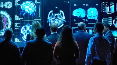
MRI (magnetic resonance imaging) uses energy from a powerful magnet to produce signals that create a series of images. These images or “slices” are analyzed by a computer to produce an image of the back and surrounding structures. MRI can help diagnose damage or disease of the spine and is particularly useful for imaging the soft tissues, such as the disks, ligaments and nerve roots in and around the spine.
Computed tomography (CT) uses a scanner to take images of the back. The images are analyzed by a computer to create reconstructed images in any plane as well as three-dimensional (3‑D) views of the back. As with MRI, CT scans help diagnose problems with the spinal canal and the surrounding tissues. CT is especially useful for looking at the bony parts of the back to detect fractures or changes from osteoarthritis. Imaging alone may not determine if your spinal stenosis requires treatment.
Signs & Symptoms of Spinal Stenosis
Symptoms of spinal stenosis may develop when the spaces within the spine narrow, most often in the lower back and neck. The narrowing creates pressure on the spine and related structures, causing symptoms. For most people, symptoms develop and progress slowly over a period of time, and some people may not have any symptoms.
The symptoms you experience depend on the location of the narrowing in your spine. Symptoms of spinal stenosis in the lower back can include:
- Pain in the lower back
- Burning pain or ache that radiates down the buttocks and into the legs, that typically worsens with standing or walking and gets better with leaning forward (flexion)
- Numbness, tingling or cramping in the legs and feet; these may become more pronounced during standing or walking
- Weakness in the legs and feet
Symptoms of spinal stenosis in the neck may include:
- Neck pain
- Numbness or tingling that radiates down the arms into the hand
- Weakness in a hand, arm or fingers
Walking, standing or extending the lumbar area of the spine can cause symptoms to worsen. Sitting or flexing the lower back or neck may relieve symptoms. The flexed position “opens up” the spinal column, enlarging the spaces between vertebrae at the back of the spine.
People with more severe stenosis may have problems with:
- Bowel function
- Bladder function
- Sexual function
Diagnosing Spinal Stenosis
Doctors use a variety of tools to diagnose spinal stenosis and rule out other conditions. Your doctor will ask about your medical and family history. This helps to determine if an injury, aging or an underlying condition is the cause of your symptoms. Some questions your doctor may ask:
- Where is the exact location of your pain, and does the pain radiate anywhere (e.g., back, legs, feet)?
- Can you describe your symptoms (e.g., aching, burning, tingling)?
- When did your symptoms begin?
Physical Exam
Your doctor will likely perform a physical exam, which may check:
- The limits of your ability to move
- If you have pain or symptoms when you hyperextend the spine (bend backwards) or if the pain improves when you bend forward
- Neurological function, such as sensation, muscle strength and reflexes in the arms and legs
- Your balance
- How you walk
Imaging tests such as X-rays show bones and can help diagnose:
- Calcification
- Disc and facet joint degeneration
- Inherited conditions
- Injury such as broken bones (fractures)
- Spondylolisthesis
- Tumors
MRI (magnetic resonance imaging) uses energy from a powerful magnet to produce signals that create a series of images. These images or “slices” are analyzed by a computer to produce an image of the back and surrounding structures. MRI can help diagnose damage or disease of the spine and is particularly useful for imaging the soft tissues, such as the disks, ligaments and nerve roots in and around the spine.
Computed tomography (CT) uses a scanner to take images of the back. The images are analyzed by a computer to create reconstructed images in any plane as well as three-dimensional (3‑D) views of the back. As with MRI, CT scans help diagnose problems with the spinal canal and the surrounding tissues. CT is especially useful for looking at the bony parts of the back to detect fractures or changes from osteoarthritis. Imaging alone may not determine if your spinal stenosis requires treatment.
Treating Spinal Stenosis
Doctors treat spinal stenosis with different options such as nonsurgical treatments, medications and surgical treatments.
Nonsurgical Treatments
Physical therapy to maintain motion of the spine, strengthen abdominal and back muscles, and build endurance, all of which help stabilize the spine. You may be encouraged to try slowly progressive aerobic activity, such as swimming or using exercise bicycles. In addition, your physical therapist or healthcare provider may recommend home exercises.
Sometimes a brace is used to provide some support and help you regain mobility. This approach is sometimes used for people with weak abdominal muscles or older patients with age-related changes at several levels of the spine.
Complementary and alternative treatments that may help relieve pain. Some examples include:
- Manipulation of the spine and nearby tissues. Professionals use their hands to adjust and massage the spine and muscles.
- Acupuncture, which is a Chinese practice that uses thin needles that may relieve pain in some patients.
Medications
Your doctor may prescribe one or more of the following medications to help manage the pain and inflammation caused by spinal stenosis:
- Anti-inflammatory medications to help relieve inflammation and pain
- Over-the-counter pain relievers taken by mouth or applied to the skin
- Prescription pain relievers for severe or acute pain
- Anti-inflammatory or numbing injections for pain that radiates or travels due to nerve compression or irritation
Surgical Treatments
If, after trying nonsurgical treatments and medications, you still have symptoms, your doctor may recommend meeting with a surgeon to talk about surgery. However, doctors may recommend surgery right away if you have numbness or weakness that interferes with walking, impaired bowel or bladder function, or other neurological involvement.
The decision to have surgery depends on how nonsurgical treatments have helped your symptoms, the amount of pain you feel, other diseases and conditions you may have, and your overall health. However, not everyone is a candidate for surgery, even if symptoms persist. In addition, your surgeon will review the risks and possible benefits of the surgery or procedure.
Surgeons can relieve pressure on the spinal cord and nerves, and restore spine alignment and health, by performing surgery. Possible surgeries include:
- Laminectomy: Surgery that doctors perform to treat spinal stenosis by removing the bony spurs and the bone walls of the vertebrae. This helps to open up the spinal column and remove the pressure on the nerves. Doctors may perform a discectomy during a laminectomy. A discectomy involves removing part of the herniated disk to relieve pressure on the spinal cord or nerve root. A facetectomy involves removing part or all of a facet joint to relieve pressure.
- Spinal fusion: Surgery that helps treat age-related changes to the spine and spondylolisthesis by joining two or more vertebrae in the spine that have slipped from their normal position. During this procedure, the surgeon may remove the disk between the vertebrae and uses bone grafts or metal devices to secure bones together.
- Minimally invasive surgery: Type of surgery that uses smaller incisions than standard surgery. Minimally invasive surgery may cause less scarring and damage to nearby muscles and other tissues. It can lead to less pain and faster recovery after surgery.
Removing and repairing the areas of the spine that are creating pressure usually helps decrease symptoms. Most people have less leg pain and can walk better after surgery. However, if nerves were badly damaged before surgery, there may be some remaining pain or numbness or no improvement. Also, the degenerative process may continue, and pain or limitation of activity may reappear after surgery.
Doctors who can provide treatment of spinal stenosis may be:
- Family or primary care doctors
- Neurologists, who treat disorders and diseases of the spine, brain and nerves
- Neurosurgeons, who perform surgery for disorders and diseases of spine, brain and nerves
- Orthopaedists, who treat and perform surgery for bone and joint diseases
- Pain specialists, who are physicians including anesthesiologists with specialized training in evaluation, diagnosis and treatment of different types of pain
- Physiatrists, who specialize in physical and rehabilitation medicine
- Physical therapists, who specialize in movement and strengthening muscles
- Rheumatologists, who specialize in treating musculoskeletal diseases and autoimmune disorders
Living with Spinal Stenosis
Living with spinal stenosis can be challenging. However, these self-care tips may help:
- Get regular exercise. Try to exercise at least three times a week for 30 minutes. Modify or adjust your activity and try to avoid doing things that can make the pain worse. Your healthcare provider or physical therapist may recommend specific exercises for you to do at home as well. Talk to your doctor before beginning any exercise program.
- Make adjustments in your daily routines that might trigger pain. Pace activities so you don’t overdo it.
- Use assistive devices to help give you stability when you walk around.
- Try changing your posture. Some people may find that flexing the spine can relieve some of their symptoms. Flexing opens the spaces in the spine, which takes pressure off the nerves and can help decrease pain.
- Practice healthy habits. For example, maintain a healthy weight, and if you smoke, quit.