


What Are Spinal Cord Tumors?
A tumor is a mass of abnormal cells that either form into a new growth or were present as a mass at birth (congenital). Tumors occur when something goes wrong with genes that regulate cell growth, allowing cells to grow and divide out of control. Tumors can form anywhere in the body. Spinal cord tumors form in the tissue inside the spinal cord, which is part of the central nervous system (CNS).
Depending on its type, a growing tumor may not cause any symptoms or can kill or displace healthy cells or disrupt their function. A tumor can move or press on sensitive tissue and block the flow of blood and other fluid, causing pain and inflammation. Some tumors don't cause any changes.
Tumors can be noncancerous (benign) or cancerous (malignant).
- Benign tumors can grow slowly or fast, don't spread to other parts of the body, and often can be removed surgically.
- Malignant tumors can invade surrounding tissue. Some cancerous brain tumors can be removed entirely through surgery. Some malignant tumors have edges that are hard to define, which makes it difficult for surgeons to remove the entire tumor.
Tumors can be primary or secondary.
- Primary tumors of the CNS are growths that begin in the brain or spinal cord. They can be either malignant or benign.
- Metastatic tumors, or secondary tumors, of the CNS are caused by cancer cells that break away from a primary tumor somewhere else in the body and spread to the CNS. They are more common than primary tumors of the CNS and occur more often in adults than in children.
There are more than 120 types of spinal cord tumors. Some are named by the type of normal cell they most closely resemble or by location. Spinal cord tumors are not contagious, nor, at this time, are they preventable.
Types of Spinal Cord Tumors
Spinal cord tumors are named by their location in the body, cell of origin, rate of growth, and malignancy. Some tumor types are more prevalent in children than in adults. Here is a listing of some common benign and malignant central nervous system (CNS) tumors.
Primary spinal cord tumors are rare tumors that begin in and around the spinal cord, and are often benign or noncancerous when they occur. In cases of malignant or cancerous tumors, they can potentially spread throughout the body.
All types of spinal cord tumors compress nearby blood vessels, nerve roots and the spinal cord, causing numbness, weakness and pain. These tumors, whether malignant or benign, can also affect control of the bowel and/or bladder. There are two types of spinal cord tumors, and they are classified by where they grow. Tumors that form within the nerve tissue of the spinal cord are called intramedullary tumors.
Types of intramedullary tumors include:
- Astrocytoma: These tumors have star-shaped glial cells called astrocytes, and can be low-grade or malignant. They form on the spinal cord.
- Ependymoma: The most common type of spinal cord tumor, ependymoma originates in the ependymal cells that line the central canal of the spinal cord. The ependymal cells help determine the direction of spinal cord fluid flow.
- Hemangioblastoma: These tumors involve several blood vessels and form anywhere on the spinal cord.
- Lipoma: This rare congenital tumor (present at birth) typically forms in the middle of the back in fat tissue, and ultimately presses against the spinal cord.
The other type of spinal cord tumor is the intradural-extramedullary tumor. These form in the outer layer of the spinal cord known as the dural sheath.
- Meningioma: These tumors are more common in women, and appear at the upper part of the back. They begin in the thin membranes covering the spinal cord.
- Myxopapillary Ependymoma: These tumors form in the tissue around the spinal cord.
- Neurofibroma: Neurofibromas are commonly found in people diagnosed with neurofibromatosis, a condition that causes nervous system and skin tumors. They develop in the protective covering of the spinal cord and nerves.
- Schwannoma: Schwann cells produce the insulation for the nerves. Schwannoma tumors are made of cancerous cells found outside the spinal cord, within the dura.
Causes of Spinal Cord Tumors
Researchers really don't know why primary spinal cord tumors develop. Possible causes include viruses, defective genes, exposure to certain chemicals and other hazardous materials, and immune system disorders. Sometimes central nervous system (CNS) tumors may result from specific genetic diseases, such as neurofibromatosis and tuberous sclerosis, or exposure to radiation.
The grade of a tumor may be used to tell the difference between slow-growing and fast-growing types of the tumor. The World Health Organization (WHO) tumor grades are based on how abnormal the cancer cells look under the microscope and how quickly the tumor is likely to grow and spread. Some tumors change grade as they progress, usually to a higher grade. The tumor is graded by a pathologist following a biopsy or during surgery.
- Grade I (low grade): The tumor cells look more like normal cells under a microscope and grow and spread more slowly than grade II, III and IV tumor cells. They rarely spread into nearby tissues. Grade I brain tumors may be cured if they are completely removed by surgery.
- Grade II: The tumor cells grow and spread more slowly than grade III and IV tumor cells. They may spread into nearby tissue and may recur (come back). Some tumors may become a higher-grade tumor.
- Grade III: The tumor cells tend to grow rapidly and can spread quickly into other CNS tissue. Tumor cells will look different from those in surrounding tissue.
- Grade IV: The tumor cells do not look like normal cells under a microscope and grow and spread very quickly. There may be areas of dead cells in the tumor. Grade IV tumors usually cannot be cured.
Risk Factors for Spinal Cord Tumors
Anyone can develop a spinal cord tumor, but the overall risk is very small. Risk factors for developing a spinal cord tumor include race (caucasians are more likely to develop a central nervous system tumor) and occupation. Workers in jobs that require repeated contact with ionizing radiation or certain chemicals, including those materials used in building supplies or plastics and textiles, have a greater chance of developing a spinal cord tumor.
Spinal cord tumors are common in those who have been diagnosed with disorders like Von Hippel-Lindau disease and neurofibromatosis 2. Von Hippel-Lindau disease is a multisystem disorder connected with blood vessel tumors found in the spinal cord, retina, brain as well as in the adrenal glands and kidneys. Neurofibromatosis 2 is a disorder that is hereditary and results in benign tumors on or near the nerves related to hearing.
Screening for & Preventing Spinal Cord Tumors
Most spinal cord tumors are rare, grow slowly and are unlikely to spread to other areas of the body. Spinal cord tumors can affect nerve function whether they are malignant or benign. These tumors can compress nerve tissue in the spinal cord.
Some central nervous system (CNS) tumor biomarkers have been found, such as the epithelial growth factor receptor (EGFR) gene. Researchers continue to search for additional clinical biomarkers of CNS tumors, to better assess risk from environmental toxins and other possible causes and monitor and predict the outcome of CNS tumor treatment. Identifying biomarkers may also lead to the development of new disease models and novel therapies for tumor treatment. Using biomarker tests, doctors may be able to predict how fast a tumor will grow and how you may potentially respond to chemotherapy, radiotherapy and other treatments.
Early detection is key to effective treatment, and discussing any family history of brain cancer or tumors with your providers is recommended, particularly in our parents—the source of our DNA.
Signs & Symptoms of Spinal Cord Tumors
Spinal cord tumors cause many different symptoms, which can make detection tricky. Symptoms depend on tumor type, location, size and rate of growth. Certain symptoms are quite specific because they result from damage to particular areas of the brain and spinal cord. Symptoms generally develop slowly and worsen as the tumor grows.
Common symptoms of a spinal cord tumor include:
- Pain in a specific area along the spine or that radiates from the spine to other parts of the body. The pain may be sharp or feel like burning or tingling feelings due to compression of nerves. The pain is often constant and progressive and may be severe. Back pain is a common early symptom of a spinal tumor.
- Numbness or sensory changes, which can include decreased skin sensitivity to temperature and progressive numbness or a loss of sensation, particularly in the legs.
- Motor problems and loss of muscle control, which can include muscle weakness, spasticity (in which the muscles stay stiff and contracted) and impaired bladder and/or bowel control.
Other symptoms, such as problems with bowel or bladder control or sexual dysfunction, can also occur but are less common.
Diagnosing Spinal Cord Tumors
If you are suspected of having a spinal cord tumor, your doctor (usually a neurologist, oncologist or neuro-oncologist) will perform an exam and may order a variety of tests based on your symptoms, personal and family medical history, and results of the physical exam. Once a tumor is found on diagnostic imaging studies, surgery to obtain tissue for a biopsy or removal is often recommended.
Diagnosing the type of spinal cord tumor is often difficult. Some tumor types are rare, and the molecular and genetic alterations of some tumors are not well understood. You may want to ask your primary care doctor or oncologist for a second opinion from a comprehensive cancer center or neuro-oncologist with experience treating your diagnosis or tumor type. Even a second opinion that confirms the original diagnosis can be reassuring and help you better prepare for your care and treatment.
A neurological exam can be done in your doctor's office. It assesses your movement and sensory skills, hearing and speech, reflexes, vision, coordination and balance, mental status, and changes in mood or behavior.
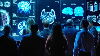
Diagnostic imaging produces extremely detailed views of structures inside the body, including tissues, organs, bones and nerves. Such imaging can confirm the diagnosis and help doctors determine the tumor's type, treatment options and, later, whether the treatment is working.
- Computed tomography (CT) scans can detect the buildup of calcium, which causes tissue to harden and develop into a tumor and can often detect hemorrhage (blood) or the development of hydrocephalus. CT scans can be done in a few minutes, so they are often used in emergency situations.
- Magnetic resonance imaging (MRI) is the gold standard for diagnosing spinal tumors and is more sensitive than a CT scan. In addition to higher resolution and better anatomic detail, MRI can provide information about blood flow (perfusion) and tumor cell density, as well as better pictures of tumors located near bone.
Usually a contrast agent (such as a dye) is injected into a vein before a CT or MRI. Many tumors become much easier to identify on the scan after the contrast is given.
- Magnetic resonance spectroscopy (MRS) can measure and analyze metabolic changes and the chemical makeup of a tissue sample.
- Magnetic resonance perfusion scans (MR perfusion) determine how fast a tumor is growing. Dye is injected into a vein, followed by an MRI scan.
- Positron emission tomography (PET) traces and measures the brain's use of glucose (sugar, used by the brain for energy) that is attached to small amounts of radioactivity and injected into the bloodstream. Because malignant tissue uses more glucose than normal tissue, it usually shows up on the scan as brighter than surrounding tissue.
- Single-photon emission computed tomography (SPECT) studies blood flow to tissue. Certain tumors grow new blood vessels to increase their supply of blood and nutrients
- Angiography (or arteriogram) can distinguish certain types of tumors that have a characteristic pattern of blood vessels and blood flow. A dye is injected into a major blood vessel, and a series of X-rays is taken as the dye flows to the brain. Often, MRI can be used to evaluate blood vessels, a procedure called MR angiography.
- Myelography is an imaging exam that can detect spinal cord inflammation or compression. These are typically performed in cases where an MRI is not possible (those with a pacemaker or a recent MR perfusion test.
- Spinal angiography tests look closely at the blood vessels that lead to and from spinal cord tumors. A catheter is inserted into a leg artery, and contrast dye is used to take pictures of the blood flow.
Laboratory and other tests:
- Testing blood, urine and other substances can provide clues about the tumor and monitor levels of therapeutic drugs.
- An electroencephalogram, or EEG, monitors brain activity through the skull (tumors can alter brain wave activity and cause seizures).
- A spinal tap (also called a lumbar puncture or CSF analysis) uses a special needle inserted into the spinal column to remove a small amount of the cerebrospinal fluid. The fluid is examined for abnormal cells or unusual levels of various molecules such as glucose and protein that suggest a brain or spinal cord tumor.
Treating Spinal Cord Tumors
A specialized team of doctors advises and assists individuals throughout treatment and rehabilitation.
- A neuro-oncologist is a neurologist or oncologist who specializes in CNS tumors.
- An oncologist is a doctor who specializes in cancer.
- A neurologist is a doctor who specializes in CNS disorders.
- A neuroradiologist is a doctor who specializes in the CNS and is trained in reading diagnostic imaging results.
- A pathologist is a clinical doctor who diagnoses diseases of tissues or cells using a variety of laboratory tests.
- A neurosurgeon is a brain or spinal cord surgeon with specialized training in removal of central nervous system tumors.
- A radiation oncologist is a doctor who specializes in using radiation to treat cancer.
Your healthcare team will recommend a treatment plan based on the tumor's location, type, size and aggressiveness, as well as your medical history, age and general health. Malignant tumors require some form of treatment, while some small benign tumors may need only monitoring. Treatment for a spinal tumor can include surgery, radiation therapy, chemotherapy, targeted therapy or a combination of treatments.
Initial treatment for a CNS tumor may involve a variety of drugs to treat or ease symptoms, including:
- anticonvulsants to treat or prevent seizures
- pain medications
- steroids or other anti-inflammatory drugs to reduce swelling and improve blood flow
- antidepressants to treat anxiety or depression that might occur following a tumor diagnosis
- anti-nausea drugs
Surgery
Surgery is usually the first treatment to both obtain tissue for diagnosis and remove as much tumor as can be done safely. Surgery may be the only treatment you need if your tumor is considered benign or low grade. Based on the type and grade (low versus high), doctors often recommend follow-up treatment, including radiation and chemotherapy, or an experimental treatment. You will be referred to the specialists above to provide guidance on this treatment.
Surgery is usually the first step in treating an accessible tumor—one that can be removed without risk of neurological damage. Many low-grade tumors and secondary (metastatic) cancerous tumors can be removed entirely. Some tumors have a clearly defined shape and can be removed more easily. Your surgeon will try removing (called resecting or excising) all or as much tumor as possible.
An inaccessible or inoperable tumor is one that cannot be removed surgically because of the risk of severe nervous system damage during the operation. In these cases, a biopsy may be performed.
A biopsy is sometimes performed to diagnose and help doctors determine how to treat a tumor. Biopsies can sometimes be performed by inserting a needle through a small hole in the body and taking a small piece of the tumor tissue. A pathologist will examine the tissue for certain changes that signal cancer and determine its stage or grade.
During surgery, some tools used in the operating room include a surgical microscope, the endoscope (a small viewing tube attached to a video camera) and miniature precision instruments that allow surgery to be performed through a small incision in the brain or spine. Other tools include:
- Intraoperative MRI, which uses a special type of MRI to provide real-time monitoring and evaluation of the surgery. Constantly updated images let doctors see how much of the tumor has been removed.
- Navigation equipment used in computer-guided, or stereotactic, neurosurgery, which gives doctors a precise, three-dimensional map of the spine as the operation progresses. A computer uses preoperative diagnostic images to reduce the risk of damage to surrounding tissue.
- Intraoperative nerve monitoring tests, which use real-time recordings of nerve cell activity to determine the role of specific nerves and to monitor brain activity as the surgery progresses. Some surgeries may be done while the individual is awake under monitored anesthesia care, rather than under general anesthesia. This allows doctors to monitor the individual's speech and motor functions as a tumor is being removed.
Radiation Therapy
Radiation therapy usually involves repeated doses of high-energy beams such as X-rays or protons to kill cancer cells or keep them from multiplying. Radiation therapy can shrink the tumor mass. It can be used to treat surgically inaccessible tumors or tumor cells that may remain following surgery.
Radiation treatment can be delivered externally, using focused beams of energy or charged particles that are directed at the tumor, or from inside the body, using a surgically implanted device. The stronger the radiation, the deeper it can penetrate to the target site. Healthy cells may also be damaged by radiation therapy, but current radiation treatment is designed to minimize injury to normal tissue.
Treatment often begins soon after surgery and may continue for several weeks. Depending on the tumor type and location, a person may be able to receive a modified form of therapy to lessen damage to healthy cells and improve the overall treatment.
Externally delivered radiation therapy poses no risk of radioactivity to the person or family and friends. Types of external radiation therapy include:
- Conventional external beam radiation, which aims a uniform dose of high-energy radiation at the tumor and surrounding tissue. It is used to treat large tumors or those that may have spread into surrounding tissue.
- Three-dimensional conformal radiotherapy (3D-CRT), which uses diagnostic imaging to prepare an accurate, computer-generated three-dimensional image of the tumor and surrounding tissue. The computer then coordinates and sends multiple beams of radiation to the tumor's exact location, sparing nearby organs and surrounding tissue.
- Intensity modulated radiation therapy (IMRT), which is similar to 3D-CRT but varies in the intensity of the hundreds of radiation beams to deliver more precise doses to the tumor or its specific areas, with less exposure to surrounding tissue.
- Hyperfractionation, which involves giving two or more smaller amounts of radiation a day instead of a larger, single dose. It can deliver more radiation to certain tumors and reduce damage to normal cells.
- Proton beam therapy, which directs a beam of high-energy protons directly at the tumor site, without spreading the radiation beyond the target. The dosing is similar to standard radiation (also called photon radiation), but proton beam radiation is best for treating tumors near important structures such as the brainstem and spinal cord. Proton beam therapy can be used as a standalone treatment or in combination with chemotherapy or as follow-up to surgery.
Radiosurgery
Radiosurgery is usually a one-time treatment using multiple, sharply focused radiation beams aimed at the spinal cord tumor from multiple angles. It does not cut into the person but, like other forms of radiation therapy, harms a tumor cell's ability to grow and divide. It is commonly used to treat surgically inaccessible tumors and may be used at the end of conventional radiation treatment.
- Linear-accelerated radiosurgery (LINAC) uses radar-like technology to prepare and fire a single beam of high-energy X-rays into the tumor. Also called high linear-energy transfer radiation, LINAC forms the beam to match the tumor's shape, avoiding surrounding tissue. A special machine that rotates around the head then fires a uniform dose of radiation into the tumor.
- Radiosurgery can be given by a number of techniques, all designed to provide a precise dose of radiation to a small area.
Side effects of radiation therapy vary from person to person and are usually temporary. They typically begin about two weeks after treatment starts and may include fatigue, nausea, vomiting, reddened or sore skin in the treated area, headache, hearing loss, problems with sleep, and hair loss (although the hair usually grows back once the treatment has stopped). Radiation therapy in young children, particularly those aged three years or younger, can cause problems with learning, processing information, thinking and growing.
There are late side effects of radiation that may occur months to years after treatment that include shrinkage (atrophy) of the spinal cord region that was treated.
Chemotherapy
Chemotherapy uses powerful drugs to kill cancer cells or stop them from growing or spreading. These drugs are usually given orally, intravenously or through a catheter or port and travel through the body to the cancerous cells. Your oncologist will recommend a treatment plan based on the type of cancer, drug(s) to be used, the frequency of administration, and the number of cycles needed. Chemotherapy is given in cycles to more effectively damage and kill cancer cells and give normal cells time to recover from any damage.
You might receive chemotherapy to shrink the tumor before surgery called neo-adjuvant therapy (a first step treatment to shrink a tumor before the primary treatment). Radiation therapy can also be given as neo-adjuvant therapy. After surgery, or radiation treatment if radiation is the primary treatment, chemotherapy could be called adjuvant therapy (treatment in addition to the primary treatment). Metronomic therapy involves continuous low-dose chemotherapy to block mechanisms that stimulate the growth of new blood vessels needed to feed the tumor.
Not all tumors are vulnerable to the same anticancer drugs, so your treatment may include a combination of drugs. Common CNS chemotherapies include temozolomide, carmustine (also called BCNU), lomustine (also called CCNU) and bevacizumab. Be sure to discuss all options with your medical team.
Side effects of chemotherapy may include hair loss, nausea, digestive problems, reduced bone marrow production and fatigue. The treatment can also harm normal cells that are growing or dividing at the same time, but these cells usually recover, and side effects often improve or stop once the treatment has ended.
Scientists at NIH and universities across the United States are exploring a variety of approaches to treat CNS tumors. These experimental approaches include boosting the immune system to better fight tumor cells, developing therapies that target the tumor cell while sparing normal cells, making improvements in radiation therapy, disabling the tumor using genes attached to viruses, and defining biomarkers that may predict the response of a CNS tumor to a particular treatment.
Targeted Therapy
Targeted therapy is a cancer treatment that uses drugs to target specific genes and proteins that are involved in tumor cell growth. This helps slow uncontrolled growth and reduce the production of tumor cells. Targeted therapies include oncogenes, growth factors and molecules aimed at blocking gene activity.
Targeted therapy uses molecularly targeted drugs that seek out the cellular changes that convert normal cells into cancer. Targeted therapies include:
- Compounds that block blood vessel growth and the flow of nutrients and oxygen to the tumor. These compounds may also hamper cell signaling and stop tumor cells from spreading elsewhere in the body.
- Developing diagnostic and therapeutic screening tools for oncogenes—transformed genes that are involved in cell growth and cause normal cells to divide uncontrollably and become malignant.
- Kinase inhibitors—proteins that block growth-signaling enzymes without harming normal cells—that may make CNS tumors more sensitive to chemotherapy.
Biological Therapy
Biological therapy (also called immunotherapy) involves enhancing the body's overall immune response to recognize and fight cancer cells. The immune system is designed to attack foreign substances in the body, but because cancer cells aren't foreign, they usually do not generate much of an immune response. Researchers are using different methods to provoke a strong immune response to tumor cells, including:
- Proteins such as interleukin and interferon and other substances that slow tumor growth
- Antibodies (proteins that are normally produced by the body to ward off bacteria and viruses) that are linked to immunotoxin drugs that seek out tumor cells and deliver their toxin, with minimal damage to surrounding normal cells
- Gene therapy, which uses a virus that can pass through the brain's protective blood-brain barrier to deliver a suicide gene to the tumor cell
- Vaccine therapy, which strengthens the immune response by inserting an antigen (a substance that triggers an immune system reaction) that the body will attack. Some vaccines attempt to target multiple antigens that the tumor may express.
Alternative & Complementary Approaches
Alternative and complementary approaches may help you or other individuals with a tumor better cope with the diagnosis and treatment. Some of these therapies, however, may be harmful if used during or after cancer treatment and should be discussed in advance with a doctor. Common approaches include nutritional and herbal supplements, vitamins, special diets, and mental or physical techniques to reduce stress.
Scientists continue to investigate ways to better understand, diagnose and treat CNS tumors. Several of today's treatments were experimental therapies only a decade ago. Clinical studies are research studies that test or observe how well medical approaches work in people. Some clinical studies test new treatments such as a new drug or medical therapy. Treatment studies help researchers learn if a new treatment is effective or less harmful than standard treatments. Studies can be considered at any point, from the time of diagnosis through recurrence.
Living with Spinal Cord Tumors
While most spinal cord tumors are benign, they may invade the nerves around the spinal cord, which can cause weakness in the limbs, loss of bowel and bladder control, and pain.
Following treatment or a medical procedure for removal of spinal cord tumors, doctors may prescribe medication for symptoms including pain. Additionally, steroid injections may reduce inflammation and muscle-spasm-related pain.
Additionally, working with a physical or occupational therapist can help with learning stretches, exercises, techniques, neuromuscular training, strengthening exercises, and help with resuming everyday activities to retain or maintain quality of life.
You may wonder how you or your loved one can help provide care for people with spinal cord tumors. Consider participating in a clinical trial so clinicians and scientists can learn more about brain and spinal tumors. Clinical research uses human volunteers to help researchers learn more about a disorder and perhaps find better ways to safely detect, treat or prevent disease.
All types of volunteers are needed—those who are healthy or may have an illness or disease—of all different ages, sexes, races and ethnicities to ensure that study results apply to as many people as possible, and that treatments will be safe and effective for everyone who will use them.
Additionally, your doctor or clinic may have a case manager who can be assigned to you who will help navigate treatment, insurance, service providers and general education about your diagnosis. Your case manager may recommend you create a binder to help keep information and treatment organized. By remaining organized, you and your loved ones can feel empowered through your treatment journey, knowing just where to find a list of medications, allergies, emergency contacts, an ongoing list of questions, your medical history, copies of imaging films, reports, lab and pathology reports—all available upon request from your caregivers. You may also wish to keep a daily journal, documenting any reactions you’re having from medications or changes in your symptoms—start times and duration are important observations to share with your treatment team.
Many patients also find value in expanding their emotional support team—mental health practitioners, friends, family and neighbors. By maintaining contact and honest communication with others, you can avoid feeling isolated and reduce anxiety, fear and depression, which are natural responses to a diagnosis.
For information about participating in clinical research, visit NIH Clinical Research Trials and You. Learn about clinical trials currently looking for people with brain and spinal tumors at Clinicaltrials.gov.