Video could not be played
Feature
The History of Einstein’s Neuroscience Department
August 20, 2024
As Einstein's Dominick P. Purpura Department of Neuroscience marks its 50th anniversary, it’s an opportune time to look back and remember the luminaries who created the department and celebrate the pioneering research by its scientists over the past five decades.
Video could not be played
When the department was founded in 1974, it immediately attracted national leaders in the field and promising young scientists who wanted to train with them.
“If neuroscience was something you wanted to do, you could not have found a better place to be than right here in the Bronx,” said Emeritus Professor Steven Walkley, D.V.M., Ph.D., a department member since 1978 (when he came to Einstein as a graduate student in Dr. Purpura’s lab) until his retirement in January.
“I was so lucky. And this place is still fantastic, thanks to the young faculty we now have and the exciting research they’re pursuing,” he added.
This article highlights a few of the many outstanding researchers who have worked in the neuroscience department during its first 50 years.
Dominick P. Purpura, M.D.
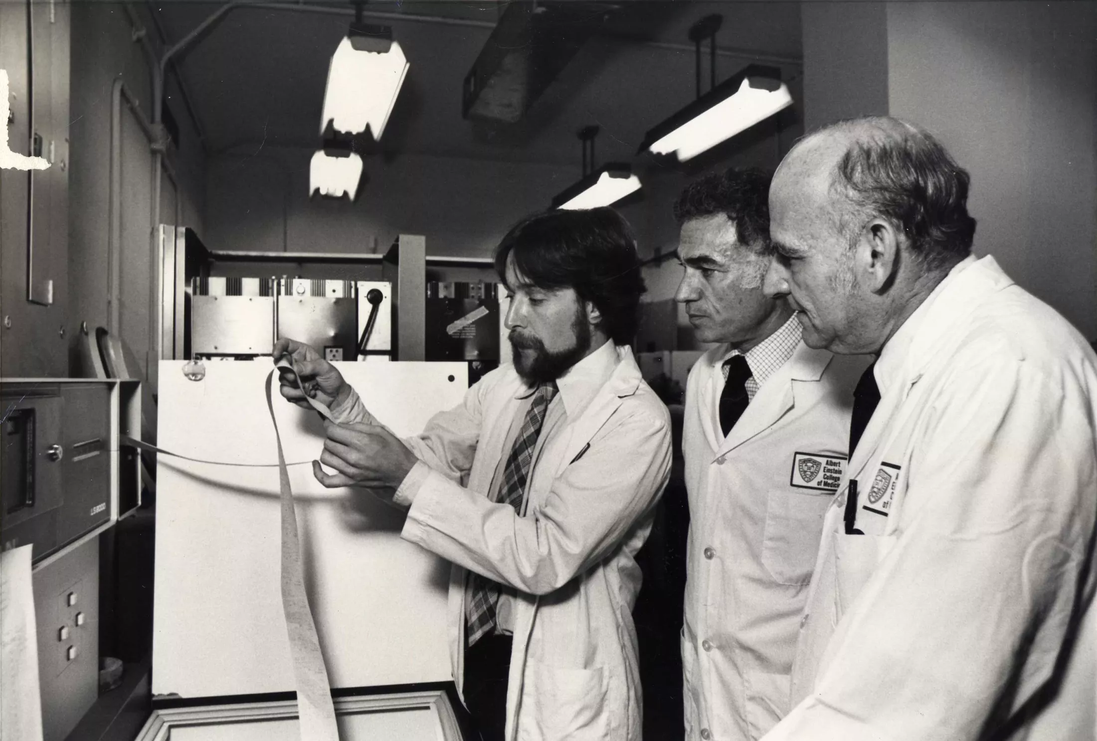
Any article on the 50th birthday of Einstein’s neuroscience department must start with Dominick Purpura, M.D. He was the founding chair of the department, which is now named in his honor, and went on to serve as Einstein’s dean from 1984 until 2006. Most of all, he was a brilliant neuroscientist. Under his direction, Einstein’s Rose F. Kennedy Center and neuroscience department became world-famous for interdisciplinary research in the brain sciences.
Video could not be played
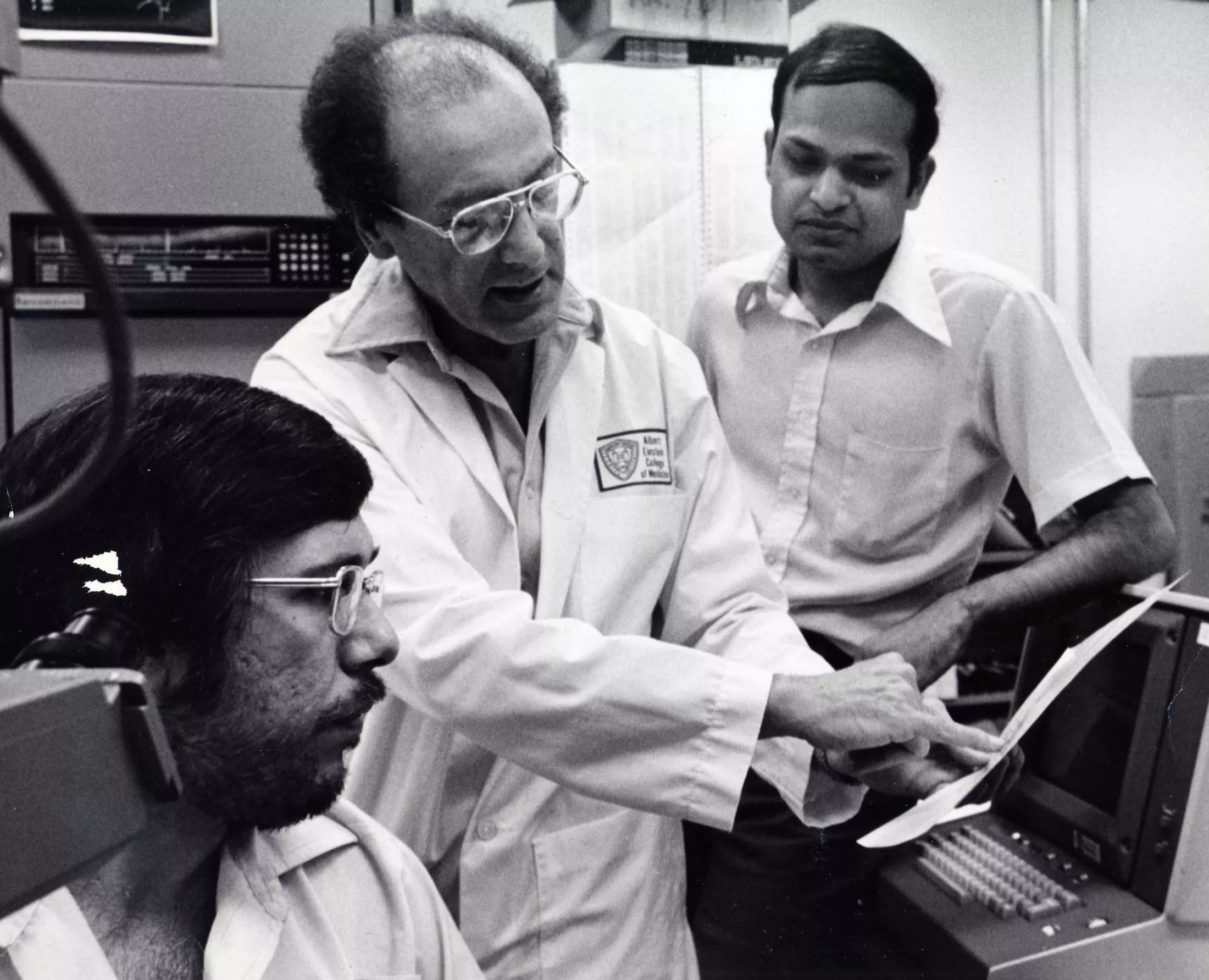
Dominick P. Purpura, M.D., center, in 1976
“Dom was Mr. Neuroscience—one of the world’s first card-carrying neuroscientists, and he created this place,” says Dr. Walkley. “Dom’s knowledge of neuroscience was encyclopedic, and he was interested in everything.”
After a decade on the faculty of Columbia University’s College of Physicians and Surgeons, Dr. Purpura joined Einstein in 1967 as professor and chair of anatomy. In 1972 he was named director of Einstein’s Rose F. Kennedy Center, which studies the causes of intellectual and developmental disabilities, and in 1974 was appointed chair of the new neuroscience department, which now occupies almost the entire nine floors of the Kennedy Center.
Dr. Purpura was widely recognized for work in several areas of neuroscience including the mechanisms of epilepsy, the origin of brain waves, fetal brain development, and—perhaps most notably—discoveries regarding structurally abnormal brain neurons in intellectual development disabilities (IDDs). His studies of an animal model of the rare IDD Tay Sachs disease and of brain tissue of affected people revealed that the loss or abnormal shape of brain neurons’ dendritic spines—tiny protrusions that contact the axons of neighboring neurons—is a hallmark of most IDDs and of cognitive developmental problems such as autism spectrum disorders.
Michael V. L. Bennett, Ph.D.
Michael Bennett, Ph.D., who succeeded Dr. Purpura as department chair, also deserves special recognition. Dr. Bennett earned an international reputation for his research on synapses: the points at which nerve impulses pass from one neuron to another. He showed that synapses are not only chemicals in nature (i.e., use acetylcholine and other chemicals to transmit messages) but that electrical synapses (commonly referred to as gap junctions) also exist.
Video could not be played
Michael Bennett, Ph.D.
“Mike’s lab—and the entire department, when he became chair—was the premier international center for gap junction electrophysiology,” said Professor David Spray, Ph.D., who came to Einstein in 1973 to work as a postdoc in the Bennett lab. “Many of the early molecular and cell biology studies of gap junctions were done here at Einstein.”
Gap junction research. The electrical synapses formed by gap junctions allow for the synchronized excitation of adjacent neurons. These junctions are found most often in invertebrate and nonmammalian nervous systems. Gap junctions are found to only a limited extent in mammalian brains; they are especially important there in synchronizing the firing of inhibitory interneurons, which are involved in all higher functions including learning, cognition, and planning.
Together with George Pappas, Ph.D., a founding member of the department, the Bennett lab discovered the structure of gap junctions by studying fish such as the electric eel, which rely on fast, electric-synapse-enabled nerve transmission for rapid escape. Through subsequent studies with colleagues and trainees, Dr. Bennett helped uncover the cellular and molecular biology of gap junctions.
Gap junctions and connexins. The “gap” between neurons is bridged by a family of some 20 proteins called connexins, which form clusters of aqueous channels that allow signaling molecules to travel between adjacent neurons. Feliksas Bukauskas, Ph.D., who often collaborated with Dr. Bennett, studied the role of connexin proteins in forming and regulating gap-junction channels; Emeritus Professor Thaddeus Bargiello, Ph.D., and Vytas Verselis, Ph.D., have studied two closely related members of the connexin gene family, Cx26 and Cx32; mutations to the gene coding for Cx26 are the most common cause of inherited deafness, and Charcot-Marie-Tooth disease is caused by mutations affecting Cx32. Dr. Spray has studied many aspects of gap-junction structure and function, including how the interactions between connexins and other proteins assemble and modulate gap junctions and how glial cells affect neuron excitability.
NMDA receptors. Emerita Professor R. Suzanne Zukin, Ph.D., studied NMDA receptors—neuronal receptors that are activated by glutamate, the main excitatory neurotransmitter in the brain. NMDA receptors are believed to play a key role in forming memories. Drs. Zukin and Bennett identified the molecular mechanisms by which protein kinases control NMDA receptor function and studied how the receptors move, or “traffic,” within neurons.
Neuropathology Research
Robert Terry, M.D., Einstein’s chair of pathology from 1969, worked closely with Saul R. Korey, M.D., founding chair of neurology at Einstein, to build an internationally recognized neuropathology group. “The fourth floor of the Kennedy center housed the greatest neuropathology group in the world,” said Dr. Walkley. “For decades, everybody who became trained in neuropathology spent time working on that floor.”
Video could not be played
From left: Peter Davies, Ph.D., Robert Terry, M.D., and Robert Katzman, M.D., in 1983
Dr. Terry was a leading expert on Tay Sachs disease as well as Alzheimer’s. He began researching Alzheimer’s in 1960 and was among the first scientists to study its plaques and tangles using electron microscopy. Dr. Terry’s colleague in many of those Alzheimer’s studies was Robert Katzman, M.D. “The two Bobs,” as they were called, conducted clinical and pathological studies on elderly patients at Einstein who had shown signs of dementia and concluded that a majority actually had Alzheimer’s disease—a condition that had long been thought of as a rare form of senile dementia. Dr. Katzman’s 1976 Archives of Neurology editorial, “The Prevalence and Malignancy of Alzheimer’s Disease,” is credited with redefining Alzheimer’s disease as a major health problem.
Peter Davies, Ph.D., a neurochemist recruited to Einstein by Dr. Terry, became one of the world’s most prominent Alzheimer’s researchers. In a 1976 letter published in The Lancet, he described analyzing the cerebral cortices of recently deceased Alzheimer’s patients and finding a severe deficit in choline acetyltransferase (the enzyme needed to synthesize the neurotransmitter acetylcholine) and acetylcholine esterase (the enzyme that breaks down acetylcholine). This led to the seminal discovery that Alzheimer brains were deficient in cholinergic neurons (nerve cells that use acetylcholine to transmit messages). Since the enzyme cholinesterase breaks down acetylcholine, Dr. Davies’ discovery led to the development of the cholinesterase inhibitor, tacrine, the first Food and Drug Administration-approved drug for treating Alzheimer’s.
Dr. Davies later became involved in the controversy—still unresolved—over which is more important in causing Alzheimer’s: beta-amyloid protein or tau protein tangles. A staunch “tauist,” he published some 250 papers on the protein’s role in the disease.
Video could not be played
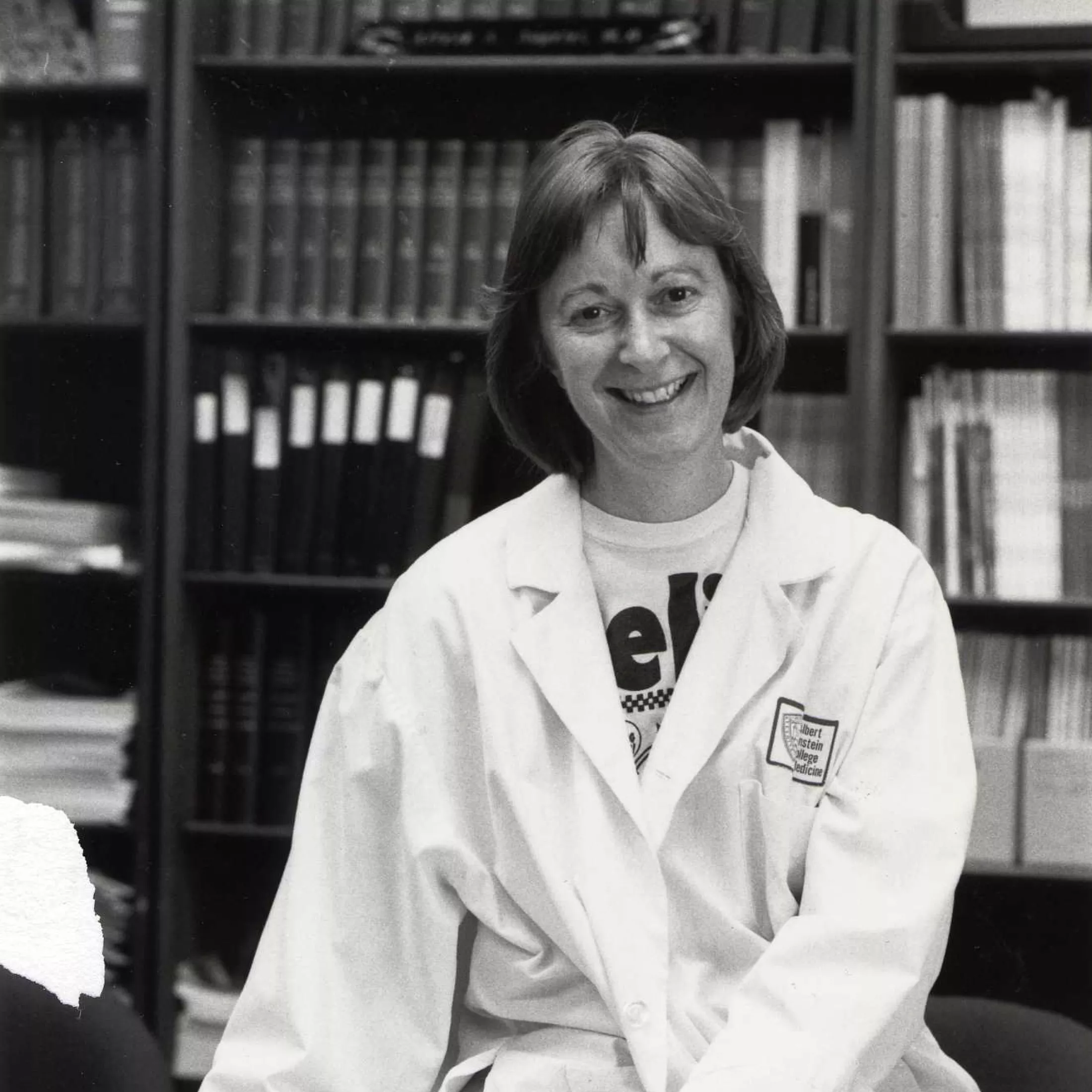
Celia Brosnan, Ph.D.
Multiple Sclerosis (MS). In the early years of the neuroscience department, the study of MS and other neuropathologies was a major area of research. Murray Bornstein, M.D., Ph.D., conducted innovative MS research using the “hanging drop” technique, which maintains neuronal or other tissue in culture so it can be studied. He also collaborated with Israel’s Weizmann Institute and Teva Pharmaceuticals to develop Copaxone, a commonly used MS treatment. Through extensive electron microscopy studies on animal models of MS, Professor Emeritus Cedric Raine, Ph.D., D.Sc., and colleagues showed that MS involves immune-system destruction of neurons’ myelin sheath. Cell culture specialist Edith Peterson, M.S., was the first person to grow myelin, the insulating sheath surrounding nerve cells—an achievement that aided MS research. Professor Emerita Celia Brosnan, Ph.D., identified the presence of lymphotoxin and tumor necrosis factor in MS brain lesions. She and fellow neurochemists William Norton, Ph.D., and Wendy Cammer, Ph.D., showed in an animal model of MS that proteolysis (degradation of proteins) is one factor contributing to the demyelination that characterizes MS.
Lysosomal storage diseases (LSDs). In Tay Sachs, Niemann-Pick, and some 50 other rare and conditions known as LSDs, inherited mutations lead to enzyme deficiencies within lysosomes—organelles that use enzymes to digest excess or broken-down cellular material. Mutated lysosomal enzymes allow toxic material to accumulate within neurons and other cells, compromising their function and causing severe intellectual disability and shortened lives.
Dr. Walkley’s research on LSDs clarified how many lysosomal diseases develop and led to therapies for two of them. In a 1994 PNAS paper involving a cat model of the LSD alpha-mannosidosis, he and colleagues reported that a bone marrow transplant works as an enzyme-replacement therapy—a finding that has led to life-saving bone-marrow transplants for a number of children with this rare disorder, which results from a deficiency of the enzyme alpha-D-mannosidase. His lab later developed two drugs, miglustat and hydroxypropyl β-cyclodextrin, that were found to slow the progression of another lysosomal disorder, Niemann-Pick type C.
The Suzukis—neuropathologist Kinuko Suzuki, M.D., and her husband, neurochemist Kunihiko Suzuki, M.D.—conducted important research on LSDs and other brain diseases, as collaborators and working independently. Dr. Kinuko Suzuki studied the LSD called late infantile GM1 gangliosidosis (caused by a deficiency of the enzyme beta-galactosidase-1). Dr. Kunihiko Suzuki studied Krabbe’s disease, an LSD caused by a deficiency of the enzyme galactosylceramidase.
Video could not be played
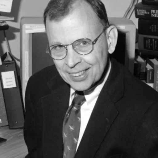
Herbert Schaumburg, M.D.
Neurotoxicology. Peter Spencer, Ph.D., and Herbert Schaumburg, M.D., collaborated to create the field of neurotoxicology and literally wrote the book on it: Experimental and Clinical Neurotoxicology. Dr. Schaumburg was an expert on peripheral neuropathies, which often result from neurotoxicity. People who used cycad seeds as a traditional food and/or medicine developed what came to be known as ALS-Parkinsonism-dementia Complex.
Electrophysiology of single neurons. At a time when culturing and studying single neurons was extremely challenging, Stanley Crain, Ph.D., (who had earlier helped develop the atomic bomb as a member of the Manhattan Project) was the first researcher to insert an electrode into a single mammalian cell in culture and record the cell’s membrane potential (i.e., electrical differencesbetween the interior and the exterior of a cell that trigger neurotransmitter release). He later studied opioid addiction and published findings regarding opioid receptors. Stephen Highstein, M.D., Ph.D., clarified the shape and physiology of individual neurons in brains of living animals using simultaneous intracellular recordings and tracer injections.
Other Areas of Neuroscience Research
Neurophysiology. Herbert Vaughan, M.D., was a major player in understanding the function of the brain’s cerebral cortex. He and colleague Joseph Arezzo, Ph.D., developed a multielectrode array for detailed electroencephalogram mapping of intracranial electrical field potentials in premature infants, yielding important insights into how the brain functions.
Video could not be played
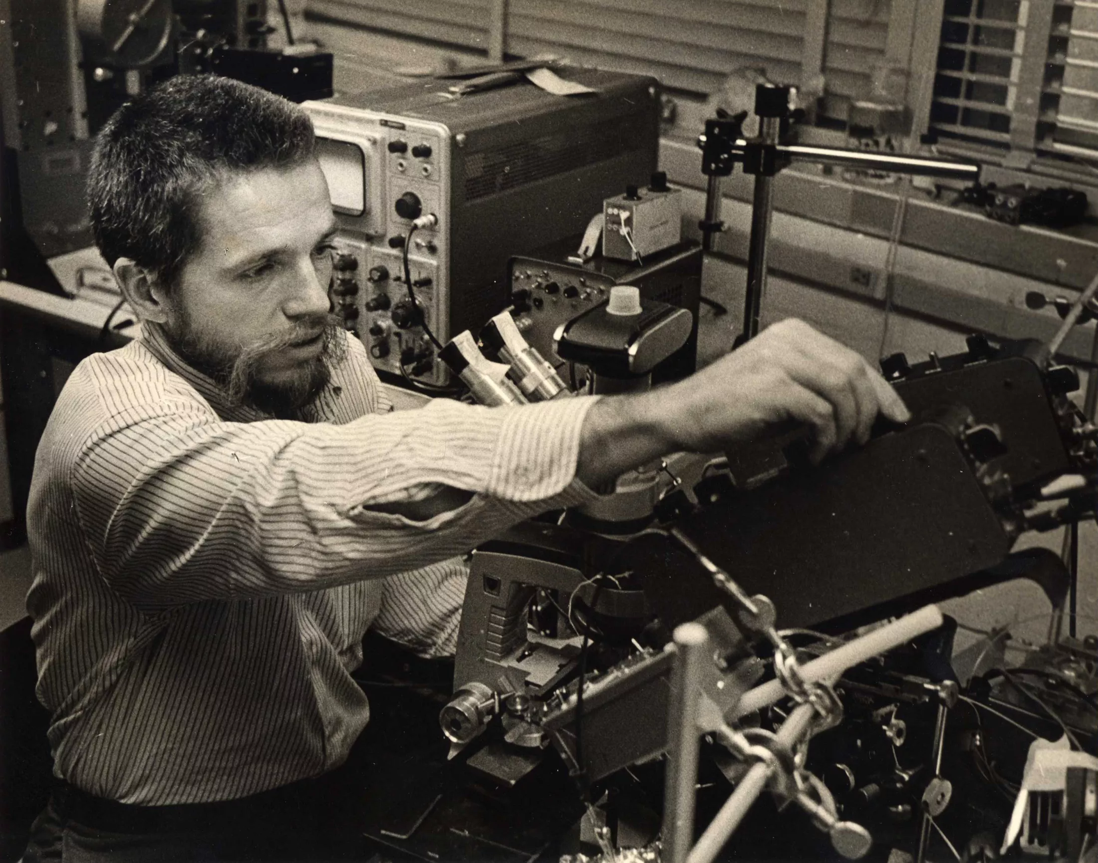
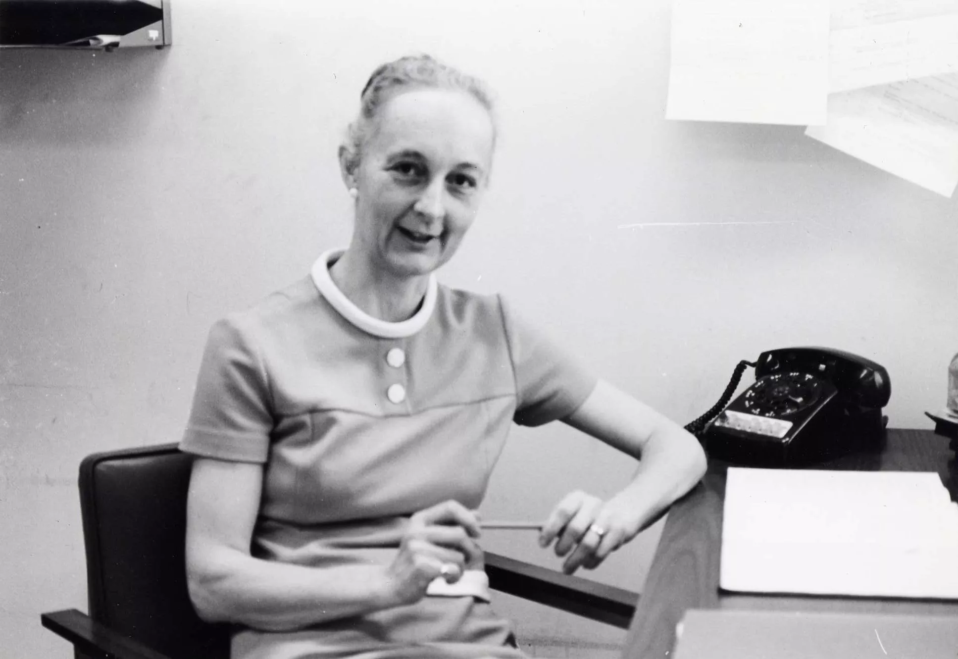
Isabelle Rapin, M.D., in 1975
Hearing. Isabelle Rapin, M.D., used techniques developed by Dr. Vaughan to record auditory evoked responses as a way to diagnose hearing loss in infants with brain damage. Dr. Rapin, who focused on communications disorders, was also a pioneer in autism research, advancing the idea that the condition was part of a broad spectrum of disorders. Robert Ruben, M.D., and Thomas Van De Water, Ph.D., conducted research on ear development, anatomy, and function, relying in part on cochleas that Dr. Van De Water grew in tissue culture.
Synaptic plasticity. Key aspects of life—how we think, feel, act, and learn—depend on information transmitted between neurons via synapses. Professor Emeritus Donald Faber, Ph.D., chair of neuroscience from 2000 to 2013, studied factors involved in regulating the strength of the synaptic connections between neurons.
Nerve regeneration. Developmental neurobiologist Pat Model, Ph.D., conducted pioneering nerve- regeneration studies in the Mexican axolotl, a salamander often used in regeneration research. Focusing on the amphibian’s Mauthner cells—a pair of large neurons in the medulla—she transplanted the neurons from the brain of one animal to another; she then used electron microscopy to determine how well the transplanted cells formed synaptic connections with their new neighbors.
Electron microscopy (EM). Scientists determine the function of a gene by knocking it out and then observing how its absence affects an animal. For the past 40 years, David Hall, Ph.D., has used EM to study more than 100 nerve-related genes in the roundworm C. elegans, an animal model that can survive virtually any gene knockout. Some of his most important findings pertain to genes that control axon guidance (the process by which neurons send out axons to their targets) and axonal transport (movement via motor protein of mitochondria and other molecular cargoes from one end of the axon to the other) by Dr. Pappas, a pioneer in the use of freeze-fracture EM, studied electric eels and other fish—research that helped reveal the structure of gap junctions as well as the dynamic nature of chemical synaptic connections and extracellular pathways in the brain.
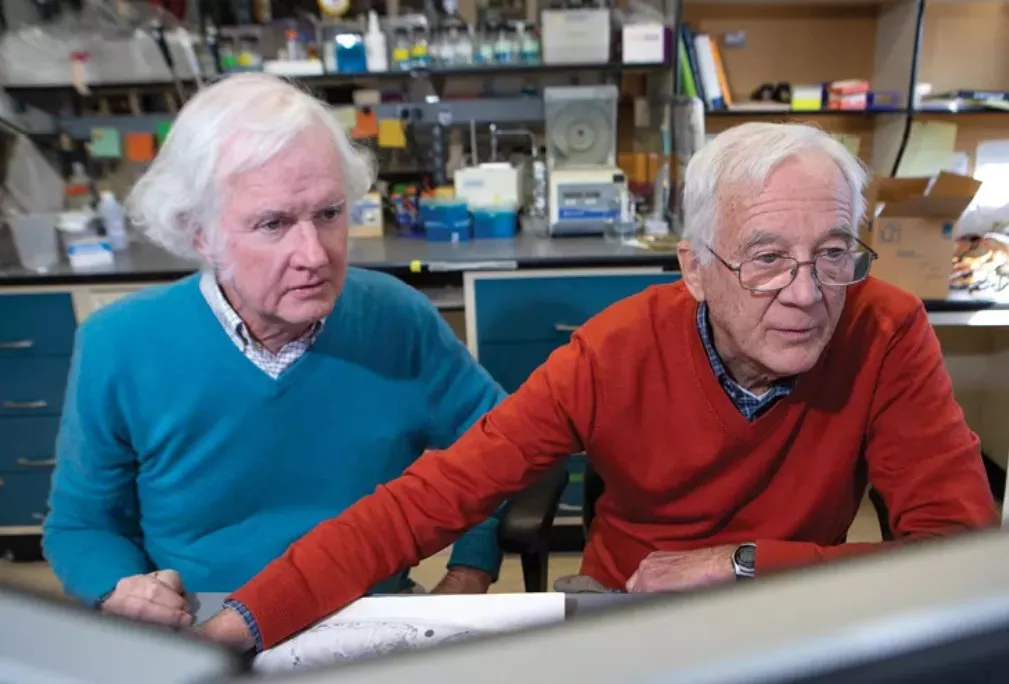
Video could not be played
David Hall, Ph.D., and Scott Emmons, Ph.D.
Connectome. Aided by Dr. Hall’s extensive collection of serial electron micrographs (the Worm Atlas) and by other Einstein colleagues, Professor Emeritus Scott Emmons, Ph.D., described the first complete wiring diagram of the nervous system of an animal, the roundworm C. elegans. The findings, published in 2019 in Nature, marked a major milestone in the field of “connectomics,” the effort to map the myriad neural connections in a brain, brain region, or simpler nervous system to find the specific nerve connections responsible for particular behaviors. Based largely on those studies, Dr. Emmons was elected to the National Academy of Sciences in 2024.
Today, a new generation of Einstein neuroscientists is continuing the outstanding research for which the department is known. Read more about Einstein faculty in Einstein’s Neuroscience Department at 50: Building on a Tradition of Excellence.